January 2004, Vol 26, No. 1 |
Update Articles
|
||||||||||||||||||||||||||||||||||||||||||||||||||
Cutaneous manifestations of viral infectionsF A Campbell, A Drummond, D T Roberts HK Pract 2004;26:31-41 Summary Viral infections are commonly seen in primary care and many do not require intervention. Comprehensive clinical history and examination should allow diagnosis but where clinical doubt exists investigation may be of value. Simple therapeutic measures are usually sufficient but more complex infections may require hospital referral for definitive diagnosis and specific intervention. 摘要 過濾性病毒感染在基層醫療中很常見,多數不需要治療。通過全面的臨床病史和體檢,可以做出明確的診斷,但若有懷疑,就要做檢查。大部份病毒感染只需要簡單的治療,但是嚴重、複雜的病例就可能要轉介到醫院以確診和給特效治療。 Introduction Viral infections are likely to present to primary care physicians and their protean manifestations may make initial diagnosis difficult. A careful clinical history and examination should provide clues which allow a definitive diagnosis to be made and appropriate therapy selected. Human papillomaviruses Papillomaviruses are a large group of small DNA viruses that infect squamous epithelium, causing cell proliferation known as warts or verrucae. The lesions can remain subclinical for long periods of time and tend to be slow growing producing no acute signs or symptoms. More than 100 different human papillomaviruse (HPV) genotypes have been identified to date and these are separated into 3 categories: cutaneous (non-genital) types such as HPV-1, -2, -3 and -4; genital-mucosal types such as HPV-6, -11, -16, and -18; and those associated with epidermodysplasia verruciformis (EV) such as HPV-5 and -8. Some genotypes appear to have malignant potential, which was first noted in the squamous carcinomas that arise in EV. Similarly the majority of cervical carcinomas contain the "high-risk" types such as 16 and 18. Non-genital warts are most common in children and young adults with an incidence of approximately 10%.1 Genital warts are generally seen in adults due to sexual transmission. In children and infants, the presence of genital warts may be a sign of abuse but equally may result from virus inoculation at birth or spread from cutaneous warts.
Warts are usually classified by their clinical location or morphology. Common warts (verruca vulgaris) can be found at any body site but are commonly found on the hands and fingers (Figure 1). Flat warts (verruca plana) are usually seen on the face, hands and legs. They are 2-4mm flat-topped papules with minimal scale. Plantar and palmar warts are thick hyperkeratotic endophytic lesions that may be painful with pressure. When these warts coalesce, they form mosaic warts (Figure 2). Anogenital warts (condylomata acuminate) vary in size and number and can form large, exophytic masses. Most patients attend their primary care physician for treatment of their warts. In the UK specialists in genitourinary medicine treat the genital lesions. There are many wart treatments available indicating that no single treatment is highly effective. The choice of treatment depends on the patient as well as the type and location of the wart. It is important to inform the patient that the wart is likely to resolve spontaneously and that no treatment is guaranteed to work. However, many children and adults wish to have treatment due to the social stigma associated with warts or to relieve pain of those on the feet or near the nails. Immunosuppressed patients usually require long-term treatment as they develop multiple warts that are difficult to eradicate. Many local treatments are available to the patient including paints, gels and salicylic acid (5-20% in yellow soft paraffin). Salicylic acid can also be combined with podophyllum resin for the treatment of plantar warts. A recent systematic review on efficacy of local treatments for warts by Gibbs et al showed topical treatments containing salicylic acid to be most efficacious with cure rates of 75%.2 Cryotherapy with liquid nitrogen is a popular treatment. It is quick and simple but can be painful and often results in blistering at the treated site. Treatment should ideally be repeated every 2-4 weeks and care needs to be taken especially around the nails and sides of the fingers where underlying structures can be damaged if used incorrectly. Cryotherapy has roughly equal efficacy to wart paints. If the wart persists despite these treatments and the patient desires further treatment several other therapies have been reported. Surgical excision, curettage or cautery is an option but recurrence rates are high. Topical immunotherapy with dinitrochlorobenzene,3 intralesional bleomycin,4 intralesional interferons,5 photodynamic therapy,6 and laser therapy7 have all be reported and may be tried for recalcitrant warts. Epidermodysplasia verruciformis Patients with EV have an increased susceptibility to cutaneous HPV infections. They present with widespread warts in childhood that involve large areas of the body or that fail to clear with treatment. They are infected with multiple types of HPV, some of which are EV specific. Half the patients inherit this condition as an autosomal recessive trait with a small number being reported as X-linked inheritance. They are at increased risk of squamous carcinoma arising in these lesions especially on sun-exposed sites and in those caused by EV HPV types. Herpes virus infection There are 8 human Herpes viruses and the resultant clinical picture is dependent on the type of infection and the immunocompetence of the host. The group comprises herpes simplex virus 1 ( HSV 1/HHV-1), herpes simplex virus 2 (HSV 2/HHV-2), Varicella zoster virus (VZV), Epstein-Barr virus (EBV), cytomegalovirus (CMV) and human herpes viruses 6, 7 and 8 (HHV-6, -7 and -8).
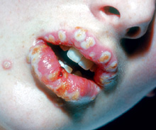
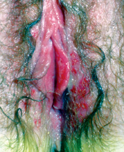
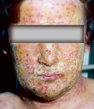
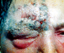
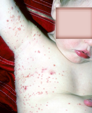
Herpes simplex (HSV 1 and 2) viruses are ubiquitous and capable of producing primary, latent and recurrent infection. HSV 1 is common in childhood and is usually spread via direct contact with a lesion or infected saliva. HSV 2 is usually sexually transmitted and consequently affects an older age group. Following contact, the virus replicates at the site of infection then travels in a retrograde direction to the dorsal root ganglion. Here it remains latent for a variable period of time. Spontaneous reactivation or stimuli such as immunosuppression, ultraviolet light exposure and physiological stress lead to the appearance of classical vesicular lesions although the original or primary infection may have passed un-noticed. Primary HSV infection in symptomatic individuals produces non-specific malaise and adenopathy, and local tenderness or burning may precede the appearance of the vesicles. Typically these have an erythematous base and are umbilicated and painful. They progress to pustulation, erosion and crusting before disappearing over several weeks (Figure 3). Recurrent infection tends to be less severe than primary. Primary genital herpes causes extremely painful vulvo-vaginitis and may progress to involve the cervix, perineum and buttocks (Figure 4). Erosive balanitis and penile shaft lesions are seen in males although they are less likely to have systemic complications such as meningitis. As in oro-labial herpes infections, recurrence tends to be less severe than primary infection but the frequency may directly correlate with the severity of the initial attack. Herpes simplex viruses may complicate atopic eczema, causing "eczema herpeticum" (Figure 5). Patients with underlying skin disorders such as burns, bullous pemphigoid or cutaneous lymphoma may also be vulnerable. Solitary herpetic lesions, "herpetic whitlows" may be seen on the digits of dentists or oral hygienists due to contact with infected patients while "herpes gladiatorum" results from direct transmission during contact sports. Herpes simplex may affect the eye causing kerato-conjunctivitis, corneal ulceration and blindness. Herpes encephalitis may cause neurological deficit or death. Neonatal herpes is more likely to arise from contact with a primary rather than a recurrent infection during pregnancy but delivery should be planned accordingly to minimise risk to the infant. Infection may be confirmed by viral culture, electron microscopy or polymerase chain reaction (PCR) which is considered to be the most accurate. Such confirmation may be required if the clinical picture is atypical and the differential diagnosis will include aphthous stomatitis or Stevens-Johnson syndrome in oral cases and syphilis or granuloma inguinale in genital infections. Simple measures will help to reduce the spread of HSV. Separate utensils and cups will minimise oral transmission and condoms are useful in genital infection provided all lesions are covered. Anti-viral agents such as famciclovir and aciclovir are now available and are effective in reducing the duration and pain of the lesions. Oral drugs are indicated for oro-labial and genital infections whereas intravenous treatment is needed for neonates, the immuno-compromised and those with systemic complications such as encephalitis. Oral prophylaxis should be considered for those subject to frequent recurrences. Varicella zoster (HHV-3) causes varicella (chickenpox) and herpes zoster (shingles). Shingles represents reactivation of latent varicella and is usually a disease of the elderly or immuno-compromised. It may also occur in children who have had chickenpox within the first 12 months of life. Primary varicella is contracted by airborne droplet infection or direct contact with vesicle fluid. Following an incubation period of 14-21 days there is a short prodrome of non-specific malaise and fever after which red papules appear on the face and scalp spreading to the trunk and limbs and becoming vesicular. The extremities are often spared and the patient remains infective until the last lesion crusts over. Most cases are benign and self-limiting but in adults the rash is often more florid and fatal pneumonia may be a feature. Congenital varicella can cause eye abnormalities and limb hypoplasia and is more likely to be a problem if infection occurs during the first trimester. Immuno-suppressed patients often have extensive atypical haemorrhagic or purpuric lesions as well as visceral involvement. In children, simple measures such as anti-pyretics and cooling topical lotions are beneficial but in adults and the immuno-suppressed, systemic antiviral drugs should be given. Varicella zoster immunoglobulin is recommended for non-immune pregnant contacts. Herpes zoster follows reactivation of varicella and may occur at any time, usually on a single dermatome. Intense pain, tingling or hyperaesthesia may precede the rash and spread across the midline is uncommon. In immuno-competent children and young adults infection causes few problems but complications become more common with increasing age and decreasing immune competence. Post-herpetic neuralgia and scarring are common and ophthalmic zoster can cause blindness (Figure 6). Immuno-suppressed patients may have atypical florid lesions and multiple dermatomes may be affected. Diagnosis of varicella will usually be evident but culture of vesicle fluid may be useful where doubt exists. Differential diagnoses include HSV, drug eruption and other viral exanthems such as coxsackie. Zoster may be mistaken for bullous impetigo or localised contact dermatitis. Herpes zoster should be treated to minimise complications and several oral preparations are available. Intravenous therapy should be used in immuno-compromised cases or in those who develop complications. Post-herpetic neuralgia may be reduced by early intervention and some success has been achieved with topical capsaicin and oral tricyclic antidepressants. The use of carbamazepine and gabapentin has also been reported. Further work is required on the benefits of varicella vaccine.8 Epstein-Barr Virus (EBV/HHV-4) is responsible for a spectrum of clinical disease including infectious mononucleosis (IM), oral hairy leukoplakia and malignancies such as nasopharyngeal carcinoma, lymphoma and Hodgkin's Disease. IM typically affects adolescents and young adults who develop fever, lymphadenopathy and a sore throat. Examination may reveal grey-white pharyngeal exudates and apart from enlarged lymph nodes there may be hepatosplenomegaly, abdominal pain and jaundice. A significant number develop a morbilliform rash which is red and sometimes haemorrhagic. All symptoms usually resolve in 2-3 weeks. Penicillin derivatives, particularly ampicillin, may precipitate an acute sensitivity eruption and this is thought to be due to infection-related IgM and IgG directed against the drug rather than to a true IgE-mediated penicillin allergy.9 Differential diagnoses should include Streptococcal tonsillitis, toxoplasmosis and lymphoma but a blood "Monospot" will confirm IM. In most cases recovery is spontaneous and treatment only supportive. Oral hairy leukoplakia causes lesions on the lateral border of the tongue and occasionally on the dorsal lingual surface or elsewhere on the oral mucosa. The ill-defined roughened white plaques are caused by benign hyperplasia of the epithelial cells and are seen predominantly in patients with HIV or in those who are immuno-suppressed for other reasons. Treatment is not usually necessary although objective improvement may be noted with retroviral therapy which in turn allows better control of the HIV. Cutaneous malignancies especially T-cell lymphomas in HIV patients may be EBV-related and are frequently poorly responsive to chemotherapy. EBV-related B-cell lymphomas have been reported in transplant patients, in patients with AIDS and in occasional patients treated with methotrexate for rheumatoid arthritis. Cytomegalovirus (CMV/HHV-5) is endemic throughout the world and infections vary in their severity according to the immuno-competence of the host. Infection is more prevalent in areas of poverty and is thought to relate to poor hygiene and overcrowding. Frequently they have associated erythema nodosum, urticaria or a morbilliform rash, worsened as in IM with the administration of ampicillin. HHV-6 gives rise to "Sixth Disease" (roseola infantum), a benign self-limiting illness usually of pre-school children. Fever is followed by a short-lived rash of circular rose-red maculo-papules sometimes with a white halo. HHV-7 is a little-understood Herpes virus which seems to be similar to HHV-6 although it is serologically distinct. It has not been clearly associated with any clinical entity and there is currently no available treatment. HHV-8 (Kaposi's Sarcoma- associated Herpes Virus/KSHV) is a latent virus found in all types of Kaposi's sarcoma (KS) and sero-prevalence for the virus can be correlated with the incidence rates of KS worldwide. HHV-8-associated diseases are thought to be a result of virus reactivation since primary infection has not yet been identified. KS was previously a rare neoplasm affecting elderly male Ashkenazy Jews or men of Mediterranean descent. In the early 1990s it was noted to be increasingly prevalent in homosexuals with AIDS and HIV positive men are now considered to have a risk 20,000-fold greater of developing KS than that of the general population.11 KS is a malignancy of vascular endothelium, classified into 4 different types - classic, HIV/AIDS-related, immunosuppression-associated and African endemic. Classic cases typically occur in elderly men as purplish lower leg plaques which slowly progress. AIDS-related lesions may initially be discrete small plaques but these evolve into florid exophytic or ulcerative lesions of the genital mucosa, oral cavity and GI tract. Progression is usual when the CD4+ count falls. Immunosuppression-related disease is similar to AIDS-related but will remit to some extent when immunosuppressive therapy is withdrawn. In African endemic disease adults or children may be affected and tumours can be highly aggressive. Differential diagnoses include angiosarcoma and pyogenic granuloma but the lesions of KS tend to be more purple in colour. There is no cure but prognosis depends to some extent on the underlying disease. Coxsackieviruses Coxsackieviruses are divided into 2 groups, A and B. Both can cause febrile exanthemic illness (often association with oral lesions), respiratory infection, aseptic meningitis and encephalitis. Group A strains cause hand, foot and mouth disease and herpangina. Group B strains cause Bornholm disease and cardiac problems. Hand, foot and mouth disease (HFMD) This is a benign self-limiting illness of childhood characterised by a vesicular eruption of the mouth and extremities. It is caused by enteroviruses; most commonly group A Coxsackieviruses. The disease occurs in epidemic form worldwide mostly in the summer and autumn months. The incubation period last 5-7 days and is followed by a non-specific prodromal illness consisting of low-grade fever, malaise and occasionally abdominal pain. In children the disease is usually mild with the cutaneous lesions dominating the clinical picture. In adults the usual presenting feature is a painful stomatitis. The skin lesions can appear simultaneously with the oral lesions or shortly thereafter. They again start as tender papules that become pearly grey vesicles with an erythematous halo. They appear on the sides or the backs of the digits, especially around the nails but may affect the palms. Foot lesions are less common than hand ones but may be found around the margin of the heels and soles. Occasionally lesions can be found on the proximal limbs, buttocks and genitalia. The diagnosis is based on the characteristic findings and no treatment is usually required. However, analgesics and topical anaesthetics can be helpful for painful oral lesions. In some large outbreaks of HFMD caused by enterovirus 71, more serious complications have been reported including CNS involvement and fatal pulmonary oedema and pulmonary haemorrhage.12 Herpangina Caused by various types of Group A Coxsackieviruses, this occurs most frequently in summer and autumn and mainly affects children. A high fever lasts 4 to 5 days and is followed by a sore throat, dysphagia, and occasionally vomiting and abdominal pain. Multiple vesicles with a vivid red areola develop on the pharynx, tonsils, uvula and soft palate. These ulcerate and then heal in 4-5 days. Treatment is symptomatic.
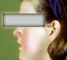
Erythema infectiosum and Parvovirus B19 infection Erythema infectiosum (fifth disease) is caused by parvovirus 19 infections and seen primarily in children. This disease is worldwide in distribution, can affect all ages and occurs throughout the year, although outbreaks tend to occur in late winter and early spring. The incubation period is 4-14 days and transmission is via aerosol spread during the viraemic phase prior to the rash appearing. Patients with Fifth disease have prodromal symptoms such as headache, coryza, malaise and low-grade fever for a few days prior to and coinciding with the rash appearing. The rash gives a characteristic "slapped cheek" appearance, which fades over 1-4 days (Figure 7). At this point, pink to erythematous macules and papules appear on the neck, trunk and extensor surfaces of the limbs and last 5 to 9 days. The rash can appear lacy or reticulate, morbilliform, confluent or annular in pattern. It is generally asymptomatic but in large outbreaks pruritus has been a major feature.13 The rash can recur for weeks to months with triggers such as sunlight, exercise, stress and bathing. Diagnosis is usually based on the clinical findings and generally no specific treatment is required or available. Papular purpuric gloves-and-socks syndrome This rare exanthem has been shown to be due to parvovirus B19. It usually affects teenagers and young adults who present with mild prodromal symptoms before a rash appears. Itchy, painful symmetrical oedema, erythema and papules of the hands and feet may be accompanied by oral lesion including erosions. Rubella (German measles) Rubella is a common, infectious viral disease which is not dangerous to most patients but which can have catastrophic effects on the developing fetus. The disease is global in distribution but immunisation programmes have helped to reduce the annual incidence and natural infection usually confers lifelong immunity. It is spread by droplet infection and the incubation period is 14-21 days. Affected individuals remain infective from the end of this period until the disappearance of the rash, typically 3-4 days later.
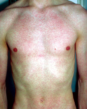
In young children there may be no prodrome but in older children and adults fever, headache, conjunctivitis and lymphadenopathy, especially occipital and post-auricular, may precede the rash by several days and resolve quickly after the spots appear. The pale reddish-pink maculo-papular eruption usually starts on the face and spreads caudally, clearing a few days later in the same order and sometimes followed by fine scaling (Figure 8). Lesions may be visible on the soft palate early on in the evolution of the rash but these are not a reliable sign. Lymphadenopathy may take weeks to resolve although local tenderness is usually short-lived. Arthropathy is uncommon in children but may be severe enough in adults to mimic rheumatoid arthritis. Although the illness is usually benign and self-limiting it is occasionally complicated by mild encephalitis with no sequelae or by thrombocytopaenic purpura which resolves in 3-4 weeks. Congenital rubella syndrome has greatly reduced in incidence as a result of childhood immunisation programmes, public awareness and effective contraception for patients who may require immunisation in adulthood. The severity of the fetal damage is dependent on the stage of pregnancy, with earlier infection causing congenital heart disease, cataract or microcephaly and later involvement causing hepatitis, myocarditis or thrombocytopaenia. Measles (Rubeola) Measles is a highly contagious viral disease of childhood which is worldwide in its distribution. The relatively asymptomatic incubation period of 10 days after exposure is followed by a 3-4 day prodrome of fever, malaise, conjunctivitis, rhinorrhoea and cough, increasing in intensity until the third phase, the rash. Typically the eruption is preceeded by "Koplik's Spots", the pathognomonic short-lived bright red lesions of the buccal mucosa which occur opposite the second molar teeth. Onset of the rash behind the ears is quickly followed by involvement of the neck, limbs and peripheries which become covered in erythematous maculo-papular lesions. These may coalesce before fading to a pale brown stain caused by capillary haemorrhage. Classical cases of measles should not create diagnostic difficulty but attenuated or atypical versions may require specific serological testing. A past history of vaccination does not preclude the illness although infection is more likely in those who have been immunised with the "killed" rather than the "live attenuated" variety. There is no specific treatment for measles but symptoms should be settled with standard anti-pyretics, oral fluids and cough suppressants where appropriate. Uncomplicated infection is usually self-limiting over a period of 10 days with no sequelae but prognosis is dependent on the host's general health and nutritional status.
Variola (Smallpox) The worldwide eradication of smallpox was announced in 1979 but recent concerns have emerged about its use as an agent of bio-terrorism. It is therefore important that primary care physicians have some knowledge of this disease and its likely mode of presentation. Infection occurs following contact with an infected human being with respiratory transmission being most likely. The incubation period is 12-13 days followed by the development of a fever, back pain and vomiting. After 3 days the typical maculopapular rash in the "swimming-trunk" area develops and is felt to be pathognomonic. The patient is not infectious during this prodromal phase. The disease may take various courses but typically the characteristic pox rash appears with papules that vesiculate and become umbilicated pustules that eventually crust and heal with scarring. Lesions vary from few to multiple and may coalesce to form large plaques. The rash is centrifugal in distribution with all the lesions at the same stage of development. The haemorrhagic form of smallpox is almost universally fatal. The overall mortality rate is approximately 30%. The diagnosis is based on the clinical findings backed up with laboratory tests including PCR detection of antigen in clinical specimens, EM, and fluorescent antibody staining of the virus. However, definite diagnosis can only be achieved by isolation of the virus in appropriate tissue culture systems. Treatment is mainly supportive although cidofovir has shown promise in animal studies.
Vaccinia This virus resulted directly from human activity in the serial propagation of viruses for human inoculation. It is used to vaccinate against smallpox, which is given to personnel working in specific scientific fields or to military personnel because of the use of variola in biological warfare. Many nations are preparing for large-scale vaccination in the event of an act of terrorism. Vaccination is not without risk as the person may develop generalised vaccinia. Progressive vaccinia may also develop if the site of inoculation fails to heal (Figure 9). This infection is clinically similar to smallpox although the lymphadenopathy is more marked. It generally occurs in children under 10 years of age and has a 17% mortality rate. Vaccination with vaccinia is protective. This disease appears only in Europe with occasional sporadic outbreaks in humans. The natural host is small rodents with most infection occurring in domestic cats. The incubation period is 2-14 days after contact and distribution of the lesions is on exposed skin. Papules rapidly become vesicular, then haemorrhagic, pustular and ulcerating. These crust over with a black eschar 1-3cm in diameter. Constitutional upset is associated with lymphangitis and lymphadenitis. Lesions heal within 3-4 weeks. Diagnosis is based on the history of contact with animals, especially cows, and the characteristic skin findings. Milker's nodules and orf should be excluded. The diagnosis can be confirmed on electron microscopy of a scraping or crust, or culture if EM is negative.
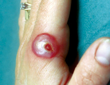
Orf This disease is widespread in sheep and goats and human lesions are due to direct inoculation of infected material. Human-to-human spread has not been reported. The disease is found most commonly in farmers and their families, veterinary surgeons and butchers. The incubation period is 5 days. A small red papule enlarges to form a flat-topped, haemorrhagic umbilicated pustule or bulla (Figure 10). The lesion is typically 2-3cm in size with a central crust and surrounding greyish white or violaceous ring, which a zone of erythema then encircles. The lesions are typically found on the fingers, hands, forearms or face and are solitary or few in number. It is associated with little or no constitutional upset and heals within 3-6 weeks. Erythema multiforme occasionally develops 10-14 days after the onset of orf. The farmers and their local physicians recognise this condition readily and generally make the diagnosis. If confirmation is needed it is best made by electron microscopy, as growth of the virus is slow and inconsistent. Milker's nodule Human infection is accidental and most common among farmers or veterinary surgeons that contract it from infected cows. The incubation period is usually 5 days after which slightly tender nodules appear on the fingers. These crust and develop surrounding erythema. The lesions heal without scarring after 4-6 weeks.
Molluscum contagiosum This is a common, benign, self-limiting infection of the skin and mucous membranes that generally affects children. This infection is less common in adults unless they are immunosuppressed, when infection can be widespread. The virus is transmitted by contact with an infected person and is seen worldwide. The lesion begins as a minute papule that enlarges to form smooth, pearly to flesh-coloured dome-shaped papules with central umbilication (Figure 11). A white, curd-like material can be expressed from the papules that vary in number from a few to hundreds, with lesions in the genitalia being frequently found in adults. Most cases resolve by 6-9 months, with or without inflammation. This infection rarely causes a diagnostic problem for the primary care physician, unless lesions are large and solitary. Treatment is not required unless requested or the patient is immunocompromised. A variety of treatments can be used including curettage and cautery, cryotherapy with liquid nitrogen, silver nitrate, imiquimod cream and cidofir. Tanapox This infection is seen mainly in Africa. Nodular lesions are associated with lymphadenopathy. The nodules ulcerate and heal with scarring after 6 weeks. Lesions are usually solitary and found on limbs. Hepatitis Hepatitis infections may have cutaneous complications. Hepatitis A, an enterovirus, is frequently accompanied by non-specific urticaria whereas patients with hepatitis B may develop erythema multiforme, cutaneous vasculitis or Gianotti-Crosti syndrome, now known to be associated with many other viral infections. Like other forms of viral hepatitis, hepatitis C (HCV) is a systemic illness but in this condition extra-hepatic manifestations, especially cutaneous ones, are more common than in other types.14 In patients with HCV infection mixed cryoglobulinaemia is a feature but the mechanism of this is unclear. Clinically it produces cutaneous vasculitis presenting as palpable purpura or frank ulceration. Other skin manifestations include lichen planus and pruritus and HCV may also be part of a triad of porphyria cutanea tarda, haemochromatosis and HCV. Oral steroids have been used with some success for cutaneous lesion but may increase viral load and create further problems. Gianotti-Crosti syndrome Gianotti-Crosti syndrome (GCS) is a cutaneous response to viral infection, originally associated with hepatitis B but now linked non-specifically to several other types of viral illness such as EBV, hepatitis A, coxsackievirus, CMV and possibly HIV. Occasional cases following immunisation against diphtheria, tetanus or polio are of uncertain significance. Similarly it is not known why the incidence peaks in the spring and summer months. The syndrome usually affects pre-school children but children of any age and occasionally adults may be affected. Clinically the rash is monomorphic with firm, non-itchy, dark red lentil-sized papules which start on the thighs and buttocks then spread to the limbs and face. Sometimes lesions become oedematous, vesicular or purpuric or coalesce to form plaques. The trunk, popliteal and antecubital fossae are usually spared. The eruption fades spontaneously in 3-4 weeks though it may last for up to 8 weeks. Constitutional upset is usually mild but soft, mobile enlarged axillary and inguinal lymph nodes may persist for weeks after the rash. Underlying infection due to hepatitis or EBV may cause derangement of liver function but this reverts to normal after a variable period. Differential diagnoses include Henoch-Schonlein purpura, lesions of which are usually larger and frankly purpuric and erythema multiforme in which lesions tend to be more obviously "targetoid". Hand, foot and mouth disease may occur in similar sites to GCS but skin lesions are normally preceded by mucous membrane involvement and palmo-plantar vesicles are characteristic. There is no specific treatment for GCS but antihistamines may be of value if itch is a problem. Human immunodeficiency virus and cutaneous disorders Human immunodeficiency virus (HIV) related skin disease ranges from exaggerated versions of common disorders to aggressive cutaneous neoplasms depending on the stage of the HIV syndrome. Initial infection is followed by assimilation of the virus into the host cells, the main target being the CD4+ lymphocyte. Where these remain at levels above 500 cells/mm3, skin problems are frequently florid versions of simple dermatoses, but when counts approach 50 cells/mm3 or less, aggressive non-healing skin lesions and malignancies become prevalent. In its earliest phase HIV infection produces a viral exanthem similar to infectious mononucleosis. This resolves spontaneously but is followed by sero-conversion to HIV positive status 6 weeks later. Common associations with HIV are herpes simplex, varicella zoster and mollusca, all of which show a progressively severe picture as the CD4+ count falls. Human papillomavirus infection may become extensive with warty lesions coalescing into large plaques. EBV-related oral hairy leukoplakia is a bad prognostic indicator heralding rapid decline and conversion to AIDS. CMV causes severe opportunistic infection and retinitis is particularly common in these patients. Non-HIV related disorders such as psoriasis may present acutely and atypically in an "inverse" distribution affecting skin flexures. Treatment can be challenging since topical agents are frequently ineffective and immunosuppressants such as methotrexate are contra-indicated. Cutaneous neoplasms such as basal cell and squamous cell carcinomata occur earlier and on non-sun exposed sites, in contrast to those seen in the normal population. Lympho-reticular malignancies are usually moderately aggressive and the association of HIV with Kaposi's sarcoma is well recognised. The clinician should be alert to the possibility of HIV infection in patients whose skin problems appear to be more florid or less responsive to treatment than the norm. Key messages
F A Campbell, MBChB, FRCP(Glasg), MRCGP, DRCOG A Drummond, BSc, MBChB, MRCP(UK)
D T Roberts, MBChB, FRCP(Glasg) Correspondence to : Dr D T Roberts, South Glasgow University Hospitals NHS Trust, Southern General Hospital, Glasgow G51 4TF, U.K. References
|
|||||||||||||||||||||||||||||||||||||||||||||||||||


