October 2006, Vol 28, No. 10 |
Update Articles
|
|||||||||||||||||||||||||||||||||||||||||||||||||||||||||||||||||||||||||||||||||||||||||||||||||||||||||||||
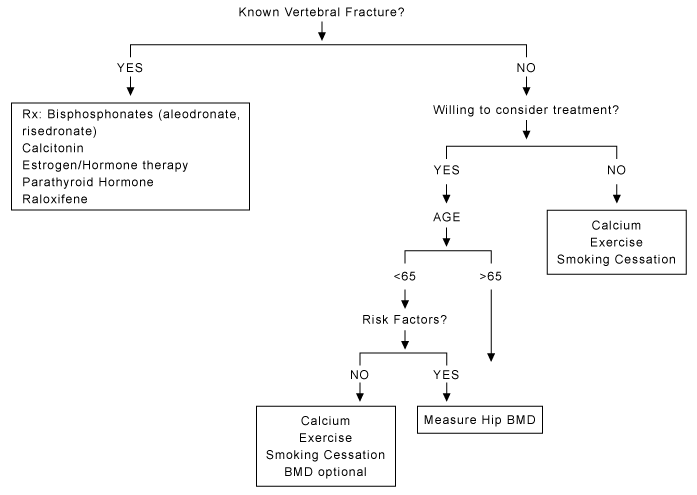
A review on osteoporosisMan-man Lam 林敏敏 HK Pract 2006;28:404-409 Summary Osteoporosis is common in postmenopausal Asian women with spinal or hip fractures being the major complications. It causes significant morbidity and mortality. Dual energy x-ray absorptiometry (DEXA) scan is the gold standard in measuring the bone mass density (BMD). Measuring BMD serves to diagnose, determine fracture risk and monitors disease progress. However, osteoporosis is still under-diagnosed. This is due to its silent presentation at the early stage, and also the difficulty to have access to the DEXA scan. According to National Osteoporosis Foundation (NOF) in United State of America, BMD measurement is indicated among those older than 65, those with risk factors, and post-menopausal women with history of bone fracture. Anti-resorptive agents or bone stimulators are treatment options. Choice of therapy should be individualized. Preventive measures are not to be under-stated. Postmenopausal women should be encouraged to take 1500 mg of supplemental daily calcium. A minimum of 30 to 60 minutes sun exposure per week is important. Regular weight-bearing exercise is to be recommended. Smoking cessation and decreased alcohol intake is to be emphasized. Fall prevention is necessary for those who are old and prone to falls. 摘要 骨質疏鬆症常見於亞裔更年期後的婦女。涉及腰椎及髖關節的骨折是它主要的併發症,因而可引起重要的後遺症。雙能量X光吸收測量儀(DEXA)是測量骨質密度指數(BMD)的標準。它能診斷骨質疏鬆,評估骨折風險,以及監察治療進度。然而骨質疏鬆症常被忽略,因為它在初期並無明顯徵狀,而且常人鮮會進行骨質密度檢查。根據美國國家骨質疏鬆症基金會指引,六十五歲或以上人仕、患骨質疏鬆的高危人仕、以及曾患骨折的更年期後婦女都需要進行骨質密度檢查。治療方面包括抗骨質吸收類及促進骨質形成類藥物。其選擇按個人情況而定。此外,亦不可忽略其他預防方法。更年期後婦女每天應補充1500毫克的鈣。每星期接觸陽光的時間應不少於30至60分鐘。定期進行負重運動是必不可少。戒煙及減少酗酒亦很重要。最後,長者及較易跌傷人仕應慎防滑倒及跌傷。 Introduction Osteoporosis is defined as low bone mass, with micro-architectural deterioration of bone tissue leading to increased bone fragility and consequent increase in bone fracture risks (Consensus 1994, WHO). Peak bone mass is reached by 25 years of age and thereafter deteriorates gradually. About one third of lifetime loss in the hip occurs before 50.1 Osteoporotic fractures are more common in whites, Asians and women than in blacks and men.2 In Hong Kong, 50% of postmenopausal women have osteoporosis.3 Women have increased bone loss over a period of 5 to 7 years after menopause, and then the rate slows down again. Osteoporosis affects 45% of women who are 50 years of age or older. It results in about 40% of lifetime fracture risk of the hip, vertebra or distal forearm. Post-menopausal women with distal radius fracture have decreased BMD and therefore nearly twice the risk of future hip fracture.4 The female-to-male fracture ratio are 7:1 for vertebral fractures, 1.5:1 for distal forearm fractures and 2:1 for hip fractures.5 Only one-third of women who sustained hip fractures are able to return to their previous living arrangements and functional abilities.6 Risk factors for osteoporosis in post-menopausal women not on HRT The major risk factors for osteoporosis are10:
Other minor risk factors are:
A retrospective cohort study performed in year 2000 found that most physicians missed the diagnosis and the chance of offering treatment in the post-menopausals.4 Why? The majority of compression fractures are asymptomatic.7 Any loss of height greater than 2.5cm should raise suspicion. Another reason of under-recognition could be attributed to poor accessibility of the BMD examination.8 BMD is interpreted in two norms, the "young normal" by T-score, and "age-matched" by Z-score.9 The WHO diagnostic category is defined using T-score, as detailed in Table 1. One standard deviation loss in bone mass, about 10 to 20% of bone loss, implies two-fold increased risk of spinal fractures or up to two-and-a-half times increased risk of hip fractures.10 BMD can be measured by DEXA, quantitative computed tomography (QCT) scan and peripheral ultrasound. Central DEXA of hip and spine is the preferred investigation for a definitive diagnosis. 9 Peripheral ultrasound assesses only the superficial bones, thus carrying certain false positives. Therefore a low peripheral bone BMD should be confirmed with central DEXA whenever possible.1
Who should be tested for BMD according to National Osteoporosis Foundation (NOF)9? Those who opt for treatment, and :
Treatment of osteoporosis The drugs used are essentially anti-resorptive agents and bone stimulators. The bone stimulators are still undergoing trials including parathyroid hormone and its related peptide analogs.12 A new agent, strontium ranelate, having both anti-resorptive and bone-forming properties is now in use. Indication for medical therapy As suggested by NOF, those at risk of or having previous fracture are to be treated.9 For all others who consider treatment, perform DEXA scan according to NOF's recommendation as mentioned above. Initiate treatment if the T-score from hip DEXA is less than -2.0, or less than -1.5 with one or more risk factors. First line therapies 1. Calcium and vitamin D The bone contains 99% of the body's calcium stores. The daily calcium requirement in post-menopausal woman and those recovering from a major fracture is 1500 mg.10 There are small but statistically significant and potentially important effects of calcium supplementation in bone loss over a two-year period.13 NOF recommends 400 IU to 800 IU per day Vitamin D in those at risk of Vitamin D deficiency. A minimum of 30 to 60 minutes of sun exposure per week is necessary to maintain adequate levels.14 The main dietary sources of vitamin D include vitamin-D-fortified milk, cereals, egg yolks, sea-water fish and liver. A recent Cochrane review indicated using vitamin D or an analogue alone was not associated with reduction in incidence of hip fractures or other non-vertebral fractures. The co-administration of calcium and vitamin D results in lower incidence of non-vertebral fractures including the hip fracture in the frail elderly (over 80 years of age).15
2. Weight-bearing exercise Exercise for more than one year is effective in slowing down bone loss.16 Aerobics, weight-bearing and resistance exercises are all effective in maintaining the BMD of the spine in postmenopausal women. Walking is effective in preventing the hip bone loss.17 Brisk walking is the best regimen for the public. To be beneficial, it should be at least 30 to 60 minutes each time and three times per week.18 There is an inverse dose-response correlation with exercise and hip fractures. Most active women show a 42% reduction in the risk of hip fractures.14 However, the benefits are lost once exercise is stopped.19 3. Avoidance of tobacco use and excessive alcohol intake Tobacco has a detrimental effect to overall health status, in addition to the effect on the skeletons. Moderate alcohol intake (i.e., one drink or less per day for women and two drinks or less per day for men)9 is associated with slightly higher bone mineral density and lower risk of fracture in the postmenopausal women. 4. Fall prevention All elderly individuals with osteoporosis should be assessed for the risk factors of fall. Side effect of drugs is a common cause of fall in elderly, other risk factors are listed in Table 2.14 Appropriate interventions can be targeted on individual risk factor. Hip protectors can be used in high risk patients.
Medical therapies 1. Bisphosphonates Bisphosphonate inhibits osteoclast-mediated bone resorption.11,12 It can be used as prevention and treatment of osteoporosis in post-menopausal women. It reduces the incidence of spinal, hip and non-spinal fractures. Studies have found that once-weekly therapy with alendronate and risedronate can prevent bone loss and fracture.20-22 Ibandronate is a potent bisphosphonate approved in Hong Kong in 2005. In the MOBILE (Monthly Oral iBandronate In LadiEs) study,23 ibandronate was administered daily at 2.5mg or once-monthly at 50+50mg (one 50-mg dose given on 2 consecutive days), 100mg or 150mg. After 2 years, greater increase in mean BMD was seen with 150mg ibandronate per month versus 2.5mg per day. It reduced the incidence of spinal fractures by about 50% over 3 years. For patients on long-term corticosteroid therapy, the rate of bone loss ranges from 0% to 13.0% per year in those who are put on at least 7.5mg per day of prednisolone.24 The bone loss is more prominent at the start of corticosteroid therapy. Bisphosphonates are effective in terms of prevention and treatment of corticosteroid-induced bone mineral loss.25,26 They should be started preferably by the time steroids are administered.27 They can be withdrawn once corticosteroid therapy is tapered off.24 Bisphosphonates are generally well tolerated,28 but some patients may experience upper gastrointestinal problems.29 Patients with oesophageal strictures, achalasia, or untreated symptomatic acid reflux should avoid bisphosphonates.14 2. Selective oestrogen receptor modulator (SERM) Raloxifene is a second generation of SERM that has been approved for the prevention and treatment of osteoporosis. It increases the bone mineral density of the vertebra by 1.28%, if one is put on a dosage of 60mg per day.1 Also it has been shown to reduce the risk of vertebral fracture to 40 or 50%. There is no reported protection concerning hip fracture.10 Raloxifene therapy results in decreased serum total and low-density lipoprotein (LDL) cholesterol levels without having any beneficial effects on serum high-density lipoprotein (HDL) cholesterol or triglyceride levels.30 Reported side effects of raloxifene are vaginitis and hot flushes.31 In addition, it showed a reduced breast cancer rate in those low risk candidates who also have post-menopausal osteoporosis after therapy for 3 years.32 3. Calcitonin Calcitonin acts by blocking osteoclastic activity. It can be administered by parental injection or intranasally. The dosage is 200IU per day. Calcitonin reduces the risk of vertebral fractures by 35%. However, it has no significant effect on the incidence of non-vertebral fractures.14 The analgesic properties of calcitonin are widely employed in pain associated with osteoporotic vertebral fracture. 4. Hormonal replacement therapy (HRT) Oestrogen decreases the incidence of both vertebral and hip fractures. In post-menopausal women oestrogen can also be used to relieve vasomotor symptoms and vulvovaginal atrophy. However, this drug has its own draw-back. In the Women's Health Initiative (WHI) study, it is found that there is a 15% increase in invasive breast cancer for women taking oestrogen plus progestogen for less than five years, and even a 53% increase for those taking it for more than five years. HRT also increased the risk of coronary heart disease, stroke and thromboembolic disease. Oestrogen, 0.625mg a day, will increase the bone mass by 2% per year. Once it is withheld, there will be a decline of bone mass up to 2% per year.10 Some studies have even suggested that there is virtually no protection benefits against fracture once oestrogen is discontinued for more than 2 to 5 years.33 It is recommended that women who discontinue oestrogen should undergo BMD assessment. If the T-score is less than -2.0, other medical therapies should be started. 5. Parathyroid hormone (PTH) PTH is approved by the FDA for the treatment of osteoporosis in post-menopausal women. The treatment significantly increases BMD at the lumbar spine and femoral neck, and it also decreases the incidence of both vertebral (65%) and non-vertebral fractures (53%) after an average of 18-months-long therapy.9 PTH causes an increased risk of osteosarcoma in rats and causes hypercalcaemia. The use of PTH should best be reserved for patients with very low BMD. The drug should be used for short duration in order to avoid possible complications.14
New agents Strontium ranelate Strontium ranelate is an orally active drug that appears to both stimulate bone formation and inhibit bone resorption. In the Treatment Of Peripheral Osteoporosis (TROPOS) study,34 strontium 2 gm/day significantly reduced the risk of vertebral and non-vertebral fractures in postmenopausal women with osteoporosis. The commonly reported adverse events were nausea, diarrhoea, headache, dermatitis and eczema. Conclusion Osteoporosis is a major health issue in the aging population. It is associated with increased risk of vertebral and non-vertebral fractures. Clinicians may miss the diagnosis due to its early silent course. There is a lack of clinical trials in the field of treatment of osteoporosis in Asian subjects. The NOF guidelines can be applied before more Asian-specific guidelines are formulated. The management plan for each patient should be individualized to achieve an effective outcome. Key messages
Man-man Lam, MBBS (HK) Correspondence to: Dr Man-man Lam, Department of Family Medicine & Primary Health Care, United Christian Hospital, Kwun Tong, Kowloon, Hong Kong. References
|
||||||||||||||||||||||||||||||||||||||||||||||||||||||||||||||||||||||||||||||||||||||||||||||||||||||||||||||