| 
                                Infective endocarditis
                                A C K Lam 林超奇 
                                HK Pract 2003;25:551-561 
                                Summary 
                                Infective endocarditis carries a high risk of morbidity and mortality. Rapid diagnosis,
                                    effective treatment and prompt recognition of the various complications are essential
                                    to a good patient outcome. Infective endocarditis often presents in an occult fashion,
                                    and its early diagnosis depends on one having a high index of clinical suspicion;
                                    especially in patients with congenital heart disease, prosthetic valves, or a previous
                                    episode of infective endocarditis. Sadly, our clinical experience shows that patients
                                    who are the sickest are often referred late for imaging and specialist care. Paradoxically,
                                    echocardiography departments are often swamped with imaging requests for patients
                                    in whom this diagnosis is unlikely.1
                             
                                摘要 
                                傳染性心內膜炎的發病和死亡的風險很高。快速的診斷,有效的治療和及早察覺併發症對治療結果有重要影響。 傳染性心內膜炎時常以不明顯的徵狀出現,早期診斷主要依靠臨床醫生的警覺性,尤其是有先天心臟病,
                                人造瓣膜和曾有傳染性心內膜炎病史的病人,患病機會很高。但實際情況, 一些嚴重的病人未能及時轉介進行造影檢查和轉介專科治療,而超聲心電圖檢查機會卻常常為染病機會不大的病人佔用了。 
 
                                Introduction 
                                Infective endocarditis (IE), or microbial infections of the endocardium is characterised
                                by fever, heart murmurs, embolic phenomena, anaemia, petechial bleeding and endocardial
                                vegetations that may result in valvular incompetence or obstruction, mycotic aneurysm
                                or myocardial abscess. The characteristic vegetative lesions are composed of a collection
                                of platelets, fibrin, microorganisms and inflammatory cells. The heart valves are
                                the most commonly involved but it could also occur on the chordae tendineae, at
                                the site of septal defect or on the mural endocardium. It has been classified as
                                "acute" or "subacute" on the basis of clinical onset, severity of the clinical manifestation
                                and the progression of the untreated disease. 
                                Hartman et al2 reviewed several major risk factors that predispose
                                to valves becoming infected. These include congenital heart disease, rheumatic heart
                                disease, degenerative heart disease, intravenous drug usage, the presence of prosthetic
                                valves, mitral valve prolapse and indwelling catheters. 
                                Incidence and epidemiology 
                                In Western Europe and the United States, the incidence of community-acquired native
                                valve endocarditis in most studies is 1.7 to 6.2 cases per 100,000 person-years.
                                Men are affected about twice as often as women.3,4 The median age of
                                onset of IE has increased from about 35 years in the pre-antibiotics era to the
                                current >50 years. This reflects increased longevity in humans with more patients
                                having degenerative valvular disease, placement of prosthetic valves, diabetes and
                                patients on long-term haemodialysis. There is an increase in nosocomial endocarditis
                                due to more cardiac surgery and other invasive procedures. Infected intravascular
                                devices give rise to at least half of these cases. There is a higher incidence of
                                right-sided endocarditis associated with intravenous drug users (IVDA) and diagnostic/therapeutic
                                procedures requiring vascular lines. Mitral-valve prolapse is a common cardiovascular
                                condition predisposing patients to IE especially in those with coexisting mitral
                                regurgitation or thickened mitral leaflets. 
                                Prosthetic-valve endocarditis (PVE) accounts for 7 to 25 percent of cases of IE
                                in most developed countries. PVE develops in 1 percent at 12 months and 2 to 3 percent
                                at 60 months after valve replacement.5,6 It is more common with aortic
                                than mitral valve prosthesis. Early-onset infections (<60 days post surgery)
                                are usually caused by coagulase-negative staphylococci. Late prosthetic-valve endocarditis
                                (>12 months post surgery) are largely community-acquired. The pathogens are usually
                                those seen in native valve endocarditis with a preponderance of viridans streptococci
                                and staphylococci, but with a high incidence of other organisms. 
                                Microbiology 
                                Overall, viridans streptococci and staphylococci account for about two-thirds of
                                all cases. In some series, staphylococci, particularly staph. aureus, have surpassed
                                viridans streptococci as the most common cause of IE. In addition, coagulase-negative
                                staphylococci, the most common pathogens in early prosthetic-valve endocarditis,
                                have also been identified as an occasional cause of native-valve endocarditis. One
                                species of community-acquired coagulase - negative staphylococcus, staph. lugdunenis
                                is commonly associated with valve destruction and the requirement for valve replacement.
                                The most common streptococci isolated from patients with endocarditis continue to
                                be Strep. sanguis, Strep. bovis, Strep. mutans and Strep. mitis. IE caused by Strep.
                                bovis is associated with colonic pathology and is more frequent in the older age
                                group. 
                                The enterococci group of organisms form part of the normal gastrointestinal flora.
                                They are more resistant to antibiotics and are more virulent then viridans streptococci.
                                There has been an increase in enterococcal endocarditis in the past decade, particularly
                                in the elderly. The most common species causing endocarditis is
                                 and occasionally  . Most cases are community-acquired
                                but the infection can be acquired in hospital post urological instrumentation. 
                                The HACEK group of organisms are fastidious slow growing species that are oropharyngeal
                                commensals and have a predilection for heart valves. The group consists of Haemophilus
                                aphrophilus/paraphrophilus, Actinobacillus actinomycetemcomitans, Cardiobacterium
                                hominis, Eikenella corrodens and Kingella kingae. Their presence in blood cultures
                                is nearly always synonymous with HACEK endocarditis. HACEK organisms endocarditis
                                tends to present as large vegetations in native valve, which might be the result
                                of prolonged illness and delay in diagnosis. 
                                For other unusually encountered organisms, the approach to the patient with an apparent
                                blood culture-negative IE, Bayer et al7 has written an excellent
                                review and discussion. 
                                Symptoms and signs 
                                Subacute bacterial endocarditis (SBE) has an insidious onset with low grade fever,
                                fatiguability, weight loss, night sweats, chills, arthralgias and valvular insufficiency.
                                Embolism of vegetations may produce cerebral vascular accident, myocardial infarction,
                                renal involvement with flank pain and haematuria, abdominal pain or acute arterial
                                insufficiency in the peripheral extremity. Physical signs include pallor, fever,
                                new or changing murmurs, tachycardia, petechiae over the upper trunk, conjunctiva,
                                mucous membranes and distal extremities, painful erythematous subcutaneous nodules
                                about the tips of the digits (Osler nodes), splinter haemorrhages under the nails,
                                non-tender erythematous haemorrhagic or pustular lesions often on the palms or soles
                                (Janeway's lesions), haemorrhagic-retinal lesions (Roth's spots - round or oval
                                lesions with small white centres). 
                                With prolonged infection, splenomegaly or clubbing of the fingers and toes may also
                                be present. Haematuria and proteinuria may result from embolic infarction of the
                                kidney or diffuse glomerulonephritis due to immune complex deposition. Neurological
                                involvement includes transient ischaemic attacks, toxic encephalopathy, brain abscess
                                and subarachnoid haemorrhage from rupture of a mycotic aneurysm. 
                                In acute bacterial endocarditis (ABE), the symptoms and signs are similar but the
                                pace of onset of disease is more rapid. ABE is characterised by the presence of
                                high fever, rapid valvular destruction, valvular ring abscesses, septic emboli,
                                toxic appearance and shock may occur. 
                                Prosthetic valvular endocarditis often results in valvular ring abscesses, obstructive
                                vegetations, myocardial abscesses, mycotic aneurysms which can present as valvular
                                obstruction, dehiscence and cardiac conduction disturbances as well as the usual
                                symptoms of SBE or ABE. The high frequency of invasive infection in prosthetic -
                                valve endocarditis results in a higher rate of new or changing murmurs, and of congestive
                                heart failure. Right-sided endocarditis is characterised by septic phlebitis, fever,
                                pleurisy, haemoptysis, septic pulmonary infarction, and tricuspid regurgitation. 
                                Diagnosis 
                                The diagnosis of IE requires the combination of clinical, laboratory, and echocardiographic
                                data. Blood culture is the most important laboratory investigation in the diagnosis
                                of endocarditis. Isolation of the pathogen enables an effective antibiotic treatment
                                regimen to be devised. Blood cultures should be taken prior to any antibiotic treatment
                                is contemplated. If antibiotics have already been given and the patient is clinically
                                stable, delaying empirical therapy for a few days and obtaining additional blood
                                cultures should be considered. It is conventional to take three to five sets of
                                blood cultures within 24 hours to isolate the aetiologic agent and to eliminate
                                the relevance of skin contaminants. 
                                Other abnormal blood results which might be present include an elevated erythrocyte
                                sedimentation rate and
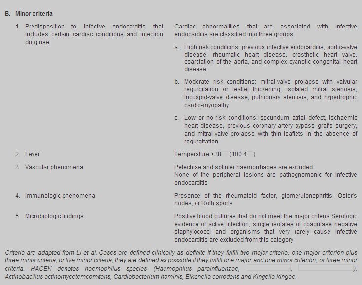
                                 protein, normochromic normocytic anaemia, leukocytosis, hypergammaglobulinaemia,
                                low serum complement, false-positive rheumatoid factor and circulating immune complexes.
                                When haematuria is present, microscopic examination for the red cell casts in a
                                centrifuged specimen would clinch the diagnosis for glomerulonephritis. For negative
                                blood cultures, serum antibodies are used to diagnose Coxiella burnetii (Q fever),
                                Bartonella and Chlamydia endocarditis. ECG evidence of new atrioventricular, fascicular,
                                or bundle-branch block would suggests perivalvular invasion in aortic - valve endocarditis. 
                                The Duke criteria 
                                In 1994 Durack8 and his colleagues introduced "the Duke Criteria" (Table
                                    1). Major criteria include: (1) a positive blood culture from microorganism
                                that typically causes infective endocarditis from two separate blood cultures; and
                                (2) evidence of endocardial involvent documented by echocardiography (definite vegetation,
                                myocardial abscess, or new partial dehiscence of a prosthetic valve) or development
                                of a new regurgitant murmur. Minor criteria include: (1) the presence of a predisposing
                                condition, (2) fever >38 , (3)
                                embolic disease, (4) immunologic phenomena (glomerulonephritis, Osler's nodes, Roth's
                                spots, rheumatoid factor), (5) positive blood cultures but not meeting the major
                                criteria and (6) a positive echocardiogram but not meeting the major criteria. A
                                definite diagnosis can be made with 80% accuracy if two major criteria, one major
                                criterion and three minor criteria, or five minor criteria are fulfilled. Modifications
                                of the Duke Criteria to increase their sensitivity have been suggested by others.9
                                These include the following additional minor criteria: the presence of newly diagnosed
                                clubbing, splenomegaly, splinter haemorrhages and petechial spots, a high erythrocyte
                                sedimentation rate or a high C-reactive protein. A single blood culture positive
                                for C. burnetti or an antiphase I IgG antibody titre >1:800 should be a major
                                criterion. 
                                   
                                Echocardiography 
                                Echocardiography is useful in diagnosis of IE. The sensitivity of transthoraic echocardiography10-12
                                is between 55% and 65%, and cannot reliably be used for confirmation of endocarditis
                                especially in patients with obesity, chronic obstructive pulmonary disease or chest-wall
                                deformities. Trans-oesophageal echocardiography12 is 90% sensitive in
                                detecting vegetative growth and is particularly useful for identifying valvular
                                ring abscesses as well as pulmonary and prosthetic valve endocarditis. 
                                Complications 
                                The clinical outcome of IE is determined by the degree of damage to the heart, the
                                location of infection (right- versus left- side, aortic versus mitral valve), whether
                                embolization has occurred from the infected site, and the immunologically mediated
                                processes. Damage to infected heart valves is common and quick with both
                                 and enterococci, but can also occur with any organism. The valvular incompetence
                                can be mild or severe and can progress even after erradication of the bacterium.
                                Aortic-valve infection is more frequently associated with congestive heart failure
                                than is mitral valve infection. Extension of IE beyond the valve annulus signifies
                                more frequent development of congestive heart failure, higher mortality and the
                                need for cardiac surgery. Extension of infection into the septum may lead to atrioventricular,
                                fascicular, or bundle-branch block. Erosion of a mycotic aneurysm of the sinus of
                                Valsalva can cause pericarditis, haemopericardium and tamponade, or fistulas to
                                the right or left ventricle. Pericarditis can also be a complication of myocardial
                                infarction due to coronary-artery embolization. 
                                Embolization commonly occurs in cerebral, myocardial, spleen, kidneys, liver, the
                                iliac or mesenteric arteries. Peripheral emboli may initiate metastatic infections
                                or send septic foci to the arterial vasa vasorum or the intraluminal vessel wall
                                causing mycotic aneurysum. Splenic abscess can be a cause of prolonged fever and
                                may cause diaphragmatic irritation with pleuritic or left shoulder pain. Up to 65%
                                of embolic events in IE involve the CNS, and neurologic complications in 20 - 40%
                                of all patients with IE. The rate of embolic events in patients with IE decreases
                                rapidly after the initiation of effective antibiotic therapy, from 13 per 1000 patient-days
                                during the first week of therapy to <1.2 per 1000 patient - days after two weeks
                                of therapy.13,14 
                                Intracranial aneurysms presentation are often variable. Some patients present with
                                no premonitory symptoms before sudden intracranial haemorrhage. Some aneurysms leak
                                slowly before rupture and produce headache and mild meningeal irritation. 
                                Fever in IE often resolves within two to three days after the start of appropriate
                                antimicrobial treatment. The most common causes of persistent fever (more than 14
                                days) are the extension of infection beyond the valve with or without myocardial
                                abscess, focal metastatic infection, drug hypersensitivity, nosocomial infection
                                or other complications of hospitalisation, such as pulmonary embolism. 
                                Treatment 
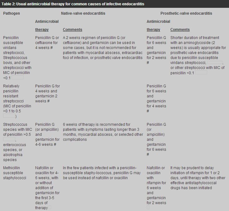
                                Treatment of the most common causes of IE is summarized in Table 2.
                                Readers are recommended to look up the Antibiotic treatment for IE due to Streptococci,
                                Enterococci, Staphylococci and HACEK microorganisms from the American Heart Association.15
                                For highly Penicillin-susceptible Viridans Streptococci or
                                 (MIC <0.1  ): treatment for 4 weeks with
                                parenteral penicillin in doses of 12 to 18 million u/24h or 2g of ceftriaxone sodium
                                in a single daily dose can achieve bacteriologic cure in up to 98% of IE patients.
                                The use of short-course (2-weeks) combination therapy with either penicillin and
                                an aminoglycoside or once-daily ceftriaxone and once-daily netilmicin has achieved
                                high bacteriologic cure in selected cases. 
                                   
                                For Penicillin-susceptible Viridans Streptococci or
                                 IE patients with prosthetic valves, a 6 weeks regimen of penicillin is recommended
                                together with gentamicin for at least the first 2 weeks. For viridans Streptococci
                                and nutritionally variant viridans streptococci (MIC >0.1 to <0.5  ) endocarditis, combination therapy with penicillin
                                and gentamicin is indicated. It is recommended that gentamicin be given for the
                                first 2 weeks of the 4 weeks course of penicillin therapy. For viridans streptococci
                                (MIC >0.5痢m/ml) endocarditis, treatment is the recommended regimen for enterococci
                                endocarditis. Treatment of enterococcal endocarditis is complicated because the
                                organisms are relatively resistant to penicillin (median MIC, 2  ), expanded-spectrum penicillins
                                or vancomycin. The increase incidence of vancomycin resistance enterococci would
                                complicate the matter even further. Nevertheless, penicillin, ampicillin or vancomycin
                                in combination with certain aminoglycoside antibiotics exert a synergistic bactericidal
                                effect on these organisms. 
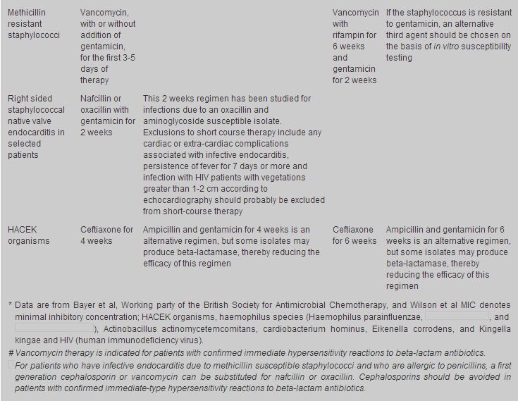
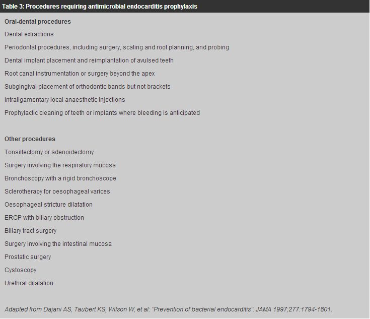
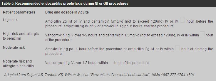
                                For methicillin-susceptible Staphylococcal endocarditis, IV nafcillin/oxacillin
                                therapy for 4 - 6 weeks together with gentamicin for the first 3 to 5 days of therapy
                                is the usual treatment. For methicillin susceptible Staphylococcal prosthetic-valve
                                endocarditis, IV nafcillin/oxacillin therapy with rifampicin for 6 weeks together
                                with gentamicin for the first 2 weeks is indicated. 
                                For methicillin resistant Staphylococcal endocarditis, the optimal antibiotic therapy
                                is vancomycin combined with rifampicin and gentamicin. Vancomycin and rifampicin
                                are administered for a minimum of 6 weeks, with gentamicin use limited to the initial
                                2 weeks of therapy. 
                                Endocarditis caused by HACEK group accounts for approximately 5% to 10% of native
                                valve endocarditis. The drugs of choice for treatment of HACEK endocarditis is ceftriaxone
                                for 4 weeks, and the duration of therapy for prosthetic valve endocarditis should
                                be 6 weeks. 
                                Uncomplicated endocarditis in selected stable, compliant patients can be managed
                                by single daily dose of ceftriaxone on an outpatient basis.16-19 This
                                flexible approach is cost effective and permits substantial cost savings in professional
                                time necessary for IV penicillin administration and the risks and inconveniences
                                inherent in any intravascular devices for four weeks. 
                                Vancomycin is an effective alternative in patients allergic to penicillins and other-lactam
                                antibiotics. Prolonged IV use of this drug may be complicated by occurrence of thrombophlebitis,
                                rash, fever, anaemia, thrombocytopenia, and (rarely) ototoxic reactions. This agent
                                should be infused over at least 1 hour to reduce the risk of the histamine-release
                                "red man" syndrome. 
                                Prevention 
                                Some cases of endocarditis occur after dental procedures or operations involving
                                the upper respiratory, genitourinary or intestinal tract. Prophylactic antibiotics
                                should be given to patients with predisposing congenital or valvular anomalies who
                                are to have any of these procedures (Tables 3,4,5). 


                                Mortality and relapse 
                                Wallace et al20 identified clinical markers, available within
                                the first 48 hours of admission that are strongly associated with in-hospital and
                                six month mortality in patients treated for infective endocarditis. 
                                They are (1) abnormal WBC <3 x 10⁹/1 or >11 x 109/1,
                                (2) serum albumin <30 gm/1, (3) creatinine level >133
                                 , (4) abnormal cardiac rhythm,
                                (5) presence of 2 major Duke criteria, (6) visible vegetation at initial echocardiography. 
                                These markers are readily available and should alert practitioners involved to seek
                                an early specialist opinions and possibly early surgery. 
                                Netzer et al21 found that independent determinants of event-free
                                survival were infection by streptococci and age <55 years. Long term survival
                                following infective endocarditis is 50% after 10 years and is predicted by: (1)
                                early surgical treatment, (2) age <55 years, (3) absence of congestive heart
                                failure, (4) initial presence of multiple="multiple" endocarditis symptoms. 
                                Mylonakis et al22 reviewed the mortality rate among patients
                                with IE and it varies according to the following factors: 
                                 
                                    causative microorganisms: 4 - 16% mortality for streptococeal viridans and bovis,
                                        15 - 25% mortality for enterococci, 25 - 47% mortality for staph. aureus, 5 - 37%
                                        mortality for Q fever, >50% mortality for P. aeruginosa, Enterobacteriaceae or fungi.presence of complications or coexisting conditions: e.g. congestive heart failure,
                                        neurologic events, renal failure or severe immunosuppression due to HIV infection.the development of perivalvular extension or a myocardial abscess.the use of combined medical and surgical therapy in appropriate patients. 
                                The overall mortality rates for both native valve and prosthetic valve endocarditis
                                remain as high as 20 - 25 percent,22 with death resulting primarily from
                                congestive heart failure and stroke. The mortality rate for intravenous drug users
                                with right-sided endocarditis are generally lower, approximately 10 - 30%, depending
                                on the size of the vegetations. 
                                Relapse of IE usually occurs within two months of discontinuation of antimicrobial
                                therapy. For native valve endocarditis, penicillin-susceptible viridans streptococcus
                                risk of recurrence is generally less than 2 percent; for enterococcus, 8 to 20 percent.
                                Among patients with IE caused by Staph. aureus, Enterobacteriacae, or fungi, treatment
                                failure often occurs during the primary course of therapy. A positive culture at
                                the time of valve - replacement surgery, particularly in patients with Staphylococcal
                                endocarditis, is a risk factor for subsequent relapse. The relapse rate in prosthetic-valve
                                endocarditis is approximately 10 to 15 percent, and relapse of infection may be
                                an indication for combined medical and surgical therapy.22 
                                Key messages 
                                 
                                    In pyrexia of unknown origin, Infective Endocarditis (IE) should be thought of even
                                        though it is not common and is often diagnosed late.The majority of IE are caused by Viridans Streptococci and Staphylococci.There is an increase in nosocomial endocarditis due to cardiac surgery and invasive
                                        procedures. Remember to ask about recent dental treatment.The risk of embolism in Streptococcus Viridans IE decreased drastically within the
                                        first 2 weeks of successful treatment.Intravenous drug user (IVDA) is now recognised as an increasing important underlying
                                        co-morbid condition for development of IE.The risk factors that predispose valves to IE are: congenital heart disease, rheumatic
                                        heart disease, IVDA, the presence of prosthetic valves, valvular stenosis/regurgitation
                                        and indwelling catheters.Uncomplicated IE can be managed by single daily dose of ceftriaxone on an outpatient
                                        basis in selected stable and compliant patients.Early clinical markers within first 48 hours with poor outcome of IE are: 
                                         
                                            abnormal W.B.C. count. low serum albumin concentration. creatinine level >133 abnormal cardiac rhythm. presence of 2 major Duke Criteria and visible vegetations. Long term survival is predicted by early surgical treatment, age <55 years, absence
                                        of congestive heart failure, and the initial presence of more symptoms of endocarditis. 
 
                                A C K Lam, MD(Canada), CCFP(Canada), FRACGP(Australia), FHKAM(Medicine)
                                Honorary Assistant Clinical Professor, Family Medicine Unit, Department of Medicine, 
                                The University of Hong Kong.
                                Honorary Associate Clinical Professor, 
                                Department of Community and Family Medicine, Department of Medicine, The Chinese
                                University of Hong Kong.
                                 
                                    Correspondence to : Dr A C K Lam, Room 1815 East Point Centre, 555 Hennessy
                                    Road, Causeway Bay, Hong Kong. 
 
                                References
                                
                                    Prendergast BD. Diagnosis of infective endocarditis. BMJ 2002;325:845-846.Borer JS, Isom OW (eds). Pathophysiology, evaluation and management of valvular
                                        heart disease. Adv Cardiol Basel, Karger, 2002;39:195-202.Berlin JA. Incidence of infective endocarditis in the Delawere Valley, 1988-1990.
                                        Am J Cardiol 1995;76:933-936.Hogevik H. Epidemiologic aspects of infective endocarditis in an urban population:
                                        a 5-year prospective study. Medicine (Baltimore) 1995;74:324-339.Agnihotri AK, McGiffin DC, Galbraith AJ, et al. The prevalence of infective endocarditis
                                        after aortic valve replacement. J Thorac Cardiovasc Surg 1995;110:1708-1720.Vlessis AA, Hovaguimian H, Jaggers J, et al. Infective endocarditis: ten-year review
                                        of medical and surgical therapy. Ann Thorac Surg 1996;61:1217-1222.Bayer AS, Bolger AF. Diagnosis and management of infective endocarditis and its
                                        complications. Circulation 1998;98:2936-2948.Durack DT, Lukes AS. The duke endocarditis service. New criteria for diagnosis of
                                        infective endocarditis: utilization of specific echocardiographic findings. Am J
                                        Med 1994;96:200-209.Li JS, Sexton DJ. Proposed modifications to the Duke Criteria for the diagnosis
                                        of infective endocarditis. Clin Infect Dis 2000;30:633-638.Mugge A, Daniel WG. Echocardiography in infective endocarditis: Reassessment of
                                        prognostic implications of vegetation size determined by the transthoracic and the
                                        transesophageal approach. J Am Coll Cardiol 1989;14:631-638.Greaves K, Mou D, Patel A, et al. Clinical criteria and the appropriate use of transthoracic
                                        echocardiography for the exclusion of infective endocarditis. Heart 2003;89:273-275.Thalme A, Nygren AT. Endocarditis: Clinical outcome and benefit of trans-oesophageal
                                        echocardiography. Scand J Infect Dis 2000;32:303-307.Steckelberg JM, Murphy JG. Emboli in infective endocarditis: The Prognostic value
                                        of echocardiography. Ann Inter Med 1991;114:635-640.Vilacosta I, Graupner C, et al. Risk of embolization after institution of antibiotic
                                        therapy for infective endocarditis. J Am Coll Cardiol 2002;39:1489-1495.Wilson W R, Karchmer AW. Antibiotic treatment of adults with infective endocarditis
                                        due to Streptococci, Enterococci, Staphylococci and HACEK Microorganisms. American
                                        Heart Association. JAMA 1995;274:1706-1713.Karchmer AW, Robert C. Single - Antibiotic therapy for streptococcal endocarditis.
                                        JAMA 1979;241:1801-1806.Lopardo G. Management of endocarditis: Outpatient parenteral antibiotic treatment
                                        in Argentina. Chemotherapy 2001:47(Supp 1):24-32.Froncioli P, Etienne J. Treatment of Streptococcal Endocarditis with a single daily
                                        dose of Ceftriaxone Sodium for 4 weeks. Efficacy and Out patient treatment feasability.
                                        JAMA 1992;267:264-267.Stamboulian D, Bonvehi P. Antibiotic management of outpatients with endocarditis
                                        due to penicillin - susceptible streptococci. Rev Infect Dis 1991;13(Supp 2):9160-9163.Wallace S M. Mortality from infective endocarditis: Clinical predictors of outcome.
                                        Heart 2002;88:53-60.Netzer ROM, Altwegg SC. Infective endocarditis: determinants of long term outcome.
                                        Heart 2002;88:61-66.Mylonakis E. Medical Progress: Infective endocarditis in adults. NEJM 2001;345:1318-1330.Ferrieri P, Gewitz MH. Unique features of infective endocarditis in childhood. Circulation
                                        2002;105:2115-2127.Dajani AS, Taubert KA. Prevention of Bacterial Endocarditis. JAMA 1997;277:1794-1801.
                                        Circulation 1997;96:358-366.Taubert KA, Dajani AS. Preventing Bacterial Endocarditis American Heart Association
                                        Guidelines. Am Fam Physician 1998:2.Journal of American Dental Association JADA Special Report on Endocarditis: Prevention,
                                        Cardiac Conditions, Bacteremia-producing Procedures, Prophylactic Regimens, Specific
                                        Situations and Circumstances and other considerations. Posted: August 1997.Nubile D, Mark J. Infective endocarditis. NEJM 2002;346:782-783.Steckelberg JM. Risk factors for infective endocarditis. Infect Dis Clin North Am
                                        1993;7:9-19.Mansur AJ, Grinberg M. Determinants of prognosis in 300 episodes of infective endocarditis.
                                        Thorac Cardiovasc Surg 1996;44:2-10.Sachdev M, Peterson GE. Imaging techniques for diagnosis of infective endocarditis.
                                        Infect Dis Clin North Am 2002;16:319-337.Von Reyn CF, Levy BS. Infective endocarditis: An analysis based on strict case definitions.
                                        Ann Intern Med 1981;94:505-518.Campbell RD, Bagshan M. Human performance and limitations in aviation. Blackwell
                                        Science, Oxford 1999.Dehart RL, Millet KC, Murphy J, eds. Fundamentals of Aerospace Medicine, Williams
                                        Dehart RL, Millet KC, Murphy J, eds. Fundamentals of Aerospace Medicine, Williams
                                        &Jagoda A. Medical Emergencies in commercial air travel. Emerg Med Clin North Am
                                        1997;15:251-260.Rosenberg CA. Emergencies in the air: Problems, management and prevention. J Emerg
                                        Med 1997;15:159-164.Highman B, Altland PD, Eagle H. Experimental bacterial endocarditis in altitude
                                        rats A.M.A., Arch Path 1954;58:241-257.Altland PD. Effect of altitude exposure on induction of streptococcal endocarditis
                                        in young and middle - aged rats. Aviat Space Environ Med 1982;53:44-48.Nakanishi K, Tajima F, Nakata Y. Tissue factor is associated with the non-bacterial
                                        thrombotic endocarditis induced by a hypobaric hypoxic environment in rats. Virchows
                                        Arch 1998;433:375-379.Hirata K, Ban T, Jinnouchi Y. Echocardiographic assessment of left ventricular function
                                        and wall motion at high altitude in normal subjects. Am J Cardiol 1991;68:1692-1697. |