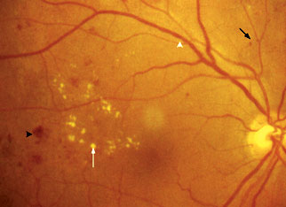
Figure 4: Microaneurysms (black arrows), retinal haemorrhages (black arrow head), hard exudates (white arrow) and venous beading (white arrow head)
| Figure 4: Microaneurysms (black arrows), retinal haemorrhages (black arrow head), hard exudates (white arrow) and venous beading (white arrow head) |
|
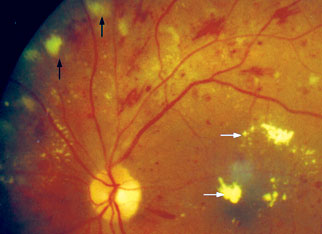
| Figure 5: Cotton wool spots (black arrows) and hard exudates (white arrows) |
 |
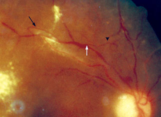
| Figure 6: Venous beading (white arrow), venous loop (black arrow) and intraretinal microvascular abnormality (black arrow head) |
 |