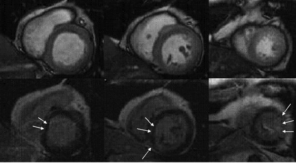
| (Top) First-pass at stress showing
concentric hypoperfusion (dark areas) in the subendocardial
layer of the left ventricle at the basal (top left panel), mid-ventricular
(top middle panel) and apex (top right panel) levels |
| (Bottom) Corresponding delayed gadolinium
hyperenhancement (white areas shown by arrows) in middle layer
of the septum at basal level (bottom left panel), in septum
and inferior wall at mid-ventricular level (bottom middle panel)
and in the lateral wall at the apex (bottom right panel)
|