September 2006, Vol 28, No. 9 |
Case Report
|
|||||||||||||||||||||||
Oral myiasis in Hong Kong - a case reportTak-shun Poon 潘德信 HK Pract 2006;28:388-393 Summary Oral myiasis - is the medical term for the invasion of parasitic larvae of flies in the oral cavity. The first published case of oral myiasis in Hong Kong was reported in 2003. The condition, caused by Chrysomya bezziana, has captured some attention amongst local people since then, especially in relation to the standard of care in institutions for the elderly. All the cases were identified among those who were debilitated, bedridden and living in homes for the aged, the adequacy of their oral care and alertness to oral myiasis by nursing care workers are questioned. We present a case of oral myiasis in a 90-year old Chinese woman, who attended our emergency department in November 2004 because of fever and upper lip swelling. We also discuss cases reported in Hong Kong, the life cycle and characteristics of the Chrysomya bezziana, and identify the predisposing factors among local patients and its intervention. 摘要 口腔蠅蛆病-是蠅的幼蟲感染人體口腔的醫學名詞。在香港自從由信氏金蠅 (Chrysomya bezziana)感染引起的口腔蠅蛆病首次在2003年被報告後,老人院的服務水平備受關注。所有得悉病例均發生在居於老人院舍的虛弱臥床年長人士。因此安老院舍對院友所提供的口腔護理和對口腔蠅蛆病的警覺性受到質疑。本文是關於一個90歲女長者的病例報告,她因上唇腫脹到急診就醫。我們亦同時討論其他曾被發表的病例,信氏金蠅的特徵和生活史,以及鑒別感染的誘因和處理方法。 Case Report We report a case of oral myiasis by Chrysomya bezziana diagnosed in our Accident & Emergency Department of Tseung Kwan O Hospital (which is a community hospital with over 400 in-patient beds and 24-hour emergency medical service). A 90-year old bed-ridden woman from a local home for the aged was brought to our department presenting with fever and upper lip swelling on 25th November 2004. The patient had multiple medical illnesses namely diabetes mellitus, hypertension, dementia and history of haemorrhagic stroke with limb contractures. She was totally dependant for her daily activities and self-care; and needed nasogastric tube feeding following the episode of her haemorrhagic stroke. Low grade fever (38.3 degree Celsius) was noted on arrival and her general condition was fair with Glasgow Coma Scale (GCS) of 7, as usual. The examination of her respiratory and cardiovascular systems revealed unremarkable findings. An oral examination showed a swollen and bleeding upper lip. Multiple dental caries and an offensive odour were noted, suggestive of very poor oral hygiene. Multiple teeth from right upper jaw were missing. The right temporomandibular joint (TMJ) was clinically subluxed which made the mouth persistently partially opened. Living maggots were seen drilling inside the mouth. More than 10 were retrieved with forceps in our department, and another 10 maggots were removed after she was admitted to the medical ward with isolation facilities immediately afterwards. Complete blood picture showed markedly elevated WBC count (22.4 x 10⁹/L) with neutrophil predominating (18.8). Liver and renal function tests were within normal range. CXR was reported as normal. Blood culture was negative and urine culture identified significant growth of Streptococcus Aerginosus. The lady was put on intravenous cefuroxime, and removal of maggots was subsequently performed every day. Totally more than 100 maggots were retrieved from her mouth. Computerized tomography of the oral cavity showed soft tissue swelling and had air density over the upper lip and adjacent gum region. Bilateral anterior dislocation of TMJ was also noted. The dental surgeon was consulted and wound debridement was done on 30th November 2004. The right palatal flap and labial flap were degloved during the procedure. Destroyed buccal soft tissue and lip skin edge were repaired and stitched. Daily oral dressing with chlorhexidine and bismuth iodoform packing was performed. Reassessment by dental surgeon later noted the buccal and palatal wound healed well with good apposition. No more maggots were seen and the lady was discharged with a surgical face mask on 13th December 2004. She was referred for follow-up by the community geriatric assessment team and for wound care by the community nursing service. The maggots sent to the hospital laboratory were identified with features compatible with those of Chrysomya bezziana. The infection control unit was informed and one living fly was found in the ward of the Hospital on 8th December 2004. Two living flies, four empty pupae and four dead flies were found on 10th December 2004.
Discussion Myiasis (Greek, myia = fly) is defined as the habit of the fly larvae (maggots) to feed on the tissues of living vertebrates.1 Myiasis occurs more frequently, although not exclusively in tropical climates like Africa, Asia, Mediterranean basin and Latin America. Disease of domestic animals is a global agricultural problem, and human cases are due to parasites that usually interact with an animal host. The appropriate travel or exposure history in a compatible clinical setting should alert the clinician to the possibility of myiasis. Although there are over 3000 species of potential agents which exist, only a limited number of species actually cause disease. Three families in the order Diptera (2-winged flies) are known to cause myiasis in humans. These are Oestridae (bot and warble flies), Calliphoridae (Blow flies), and Sarcophagidae (flesh flies). Myiasis can be classified anatomically or entomologically. The anatomic classification considers the location of parasitic infestation on the host - skin (cutaneous), nasopharyngeal, aural, ocular (ophthalmomyiasis), wound, intestinal, and urogenital myiasis. Cutaneous myiasis is the most frequently encountered clinical form. The entomologic classification places myiasis into obligatory, facultative and accidental infestations. Obligatory parasites require a living host for larval development, whereas facultative parasites might develop on either a live host or on carrion. In accidental myiasis, the eggs or larvae of Diptera are ingested in food or drink, producing moderate to severe gastrointestinal symptoms.
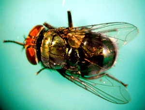
When tissues of the oral cavity are invaded by the parasitic larvae of flies, oral pathologists name this condition Oral Myiasis. Although there are some reports of oral myiasis, the classification of the larvae that cause the infestation has rarely been made. Cochliomyia hominivorax (New World screwworm) and Chrysomya bezziana, (Old World screwworms) are known to cause an obligatory oral myiasis by laying eggs directly on compromised tissue.2 New World screwworms are endemic in the southern United States, Caribbean, and most of Latin America. Old World screwworms are found in Africa, the subcontinent of India, the Arabian peninsula, and the archipelagos of Indonesia, the Philippines, and New Guinea. More mature larvae are often more invasive, readily leaving necrotic tissue for viable tissue, leading to significant local destruction, fistula formation and secondary bacterial infection. In the local case reported above, the larvae showed features characteristic of Chrysomya bezziana, belonging to the Callipharidae family. This Old World screwworm is widely distributed throughout South-East Asia, China, the Indian Subcontinent, tropical Africa, and Papua New Guinea.4 The species was first identified in Hong Kong in July 2000, when cattle infestations were noted. The first human case of Chrysomya bezziana infestation in Hong Kong was reported in 2002 as oral myiasis. According to data from Hong Kong's Department of Health, which is the central body overseeing all public health issues in Hong Kong, there were 4 reported cases of oral myiasis caused by Chrysomya bezziana in the locality, while there were 9 cases in 2003 and 8 in 2004.4 The mature larvae of C. Bezziana are notorious for their invasiveness, in the sense that, they readily leave necrotic tissue for viable tissue, and therefore causing significant local destruction, fistula formation and secondary bacterial infection. Poor oral hygiene, advanced age and high level of dependency (secondary to dementia and post-stroke limbs contracture) are identified to be common factors among the two local cases reported above. During its life cycle, adult fertile C. Bezziana female flies are usually attracted by a wound's foul odour, feed on exudates, and lay eggs in the wounded and necrotic tissues. After a day, the first larvae hatch and enter surrounding living tissues and feed for a week. The maggots will turn into adult flies in about 2 weeks.6 If living flies, empty pupae or dead flies are recovered from the ward where a known patient of oral myiasis is warded, they are likely to be associated with the index case and they should be eliminated as soon as possible. As bacterial metabolites may increase the attractiveness of infested wounds as screwworm oviposition sites, poor oral hygiene and periodontal disease may have attracted the fly as in the reported case above. Lack of self-caring ability as in dementia and cardiovascular accident cases and reduced ability of speech further deprived their power of defence to get rid of the flies. Several predisposing factors reported in other cases include diabetes mellitus, psychiatric illness, leprosy, mental subnormality, patients with an open wound, those with epilepsy who sustain trauma in the face, those who are mouth-breathers, drunkards, senile or the hemiplegic.5 The routine oral care for those who have the predisposing factors listed above (especially co-existing advanced age, high level of dependency and poor oral hygiene) are therefore particularly important and should be addressed to the concern of all nursing colleagues in the hospitals, psychiatric centres and institutions for the elderly. The treatment of oral myiasis is simple as long as the site of maggot deposition is known. Removal of the maggots and flushing the wound with normal saline or an antiseptic solution is usually curative. Recently, topical administration of 1% ivermectin (a neurotoxin) in propylene glycol for 2 hours was reported as effective and safe treatment for wound myiasis.1 However, this treatment was proven to be effective only for Cochliomyia hominivorax and its efficacy for myiasis by the same family C. Bezziana is yet to be investigated.
Family physicians should be more alert of this disease entity especially when they encounter elderly living in old aged home, who are bedridden, lacking of self-caring ability or of speech secondary to dementia or cardiovascular diseases. These patients can simply present with non-specific symptoms like fever as in our reported case or just have reduced appetite. Spending a few seconds and looking inside the mouth can spare us a lot of time and resources instead of doing blood tests recklessly for the non-specific chief complaints. Key messages
Tak-shun Poon, MBBS(HK), MRCS (Edin) Correspondence to: Dr Tak-shun Poon, Department of A&E, Tseung Kwan O Hospital, Tseung Kwan O, Kowloon, Hong Kong. References
|
||||||||||||||||||||||||