December 2008, Vol 30, No. 4 |
Update Article
|
Urology Update 1 – Urinary calculi and infectionChi-fai Ng 吳志輝 HK Pract 2008;30:199-204 Summary Urinary stone disease and urinary tract infection are
2 common urological problems. About 10% of the
population will have at least one episode of urinary stone
in their life time, and up to 50% of patients would have a
recurrence within 10 years after their initial stone
clearance. The use of non-contras t computed
tomography (NCCT) has largely replaced other imaging
as the first line investigation for ureteric colic. The
management of ureteric colic includes identifying patients
who need urgent decompression, adequate pain control
etc. The use of medical expulsive therapy, with either
alpha blocker or calcium channel blocker, can improve
the spontaneous expulsion rate of ureteric stones.
However, after the stone has been cleared, appropriate
dietary and life-style advice are important for the
prevention of stone recurrence. 摘要 泌尿道結石和泌尿道感染是泌尿科的常見病。約10%的
人一生中會有一次泌尿系結石發作;近50%的患者在初次清除
結石後10 年內會復發。非增強電腦掃描(NCCT)已被用作輸
尿管絞痛的一線檢查手段,基本取代了其它影像學方法。輸尿
管絞痛的治療包括識別需要緊急減壓的患者、充分止痛等。內
科排出療法,如使用α— 阻滯劑或鈣離子通道拮抗劑,可以
提高各種輸尿管結石的自發排出率。但在清除結石後,正確的
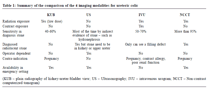
飲食及生活方式建議對預防結石復發十分重要。 Introduction As our economy and health care services become better, the life expectancy of our Hong Kong citizens is also seen to improve. Together with the increased awareness of the importance of urological problems, more and more patients are presenting themselves with various urological symptoms or diseases, including voiding problems, prostate disease, sexual dysfunction, stone disease and urological malignancies. With increasing workload in the secondary and tertiary health care system, primary care physicians will be expected to contribute more to the management of these problems. Therefore, a series of reviews on various urological conditions will be presented to update the latest developments in their management. The first review will focus on urinary stone disease and infection, followed by an update on various aspects of voiding dysfunction and finally management on urological malignancy. Urinary calculi Urinary stone disease is an ancient disease, as can be seen in the famous Hippocratic oath, which mentions this as “I will not cut for stone, even for patients in whom the disease is manifest; I will leave this operation to be performed by practitioners, specialists in this art.” About 10% of the population will have at least one episode of urinary stone in their life time. An epidemiological study on renal calculi in the Chinese population revealed that the prevalence rate was 8% and 5% in men and women respectively.1 Moreover, up to 50% of patients would have a recurrence within 10 years after their initial stone or stones were cleared. Therefore, there is a big demand for better stone care for the population, both in the acute management and preventive strategies. Management of ureteric colic Ureteric colic typically manifests as an acute, severe, and relatively constant or colicky pain starting in the renal area at around the costovertebral angle and radiating through the flank to the groin, then the scrotal area in men or labial area in women. This pain is one of the commonest presentations. However, other genito-urinary, abdominal and gynaecological emergencies can also mimic ureteric colic and therefore would impose diagnostic difficulties. The first important step in the management of ureteric colic is to confirm the diagnosis. Imaging for the diagnosis of ureteric colic While history and physical examination are essential in the diagnostic process, some form of imaging is usually necessary to confirm the presence of a stone. Although the plain kidney-ureter-bladder (KUB) x-ray is noninvasive, contrast-free and readily available in emergency departments, its accuracy in making a diagnosis of ureteric stone is limited. The reported range of sensitivity and specificity for the detection of ureteric stone are 45-62%, 67-71% respectively.2,3 Besides the limitation in diagnosing radiolucent stones, this procedure could also miss small or faint stones overlapped by bowel shadow, faecal matter and bony landmarks, especially at the sacroiliac joint. Moreover, the differentiation of a lower ureteric stone and a phlebolith could sometimes be quite difficult. Therefore, the KUB x-ray is best used in the following-up of patients with known renal calculi or as an adjunct to other imaging modalities. Ultrasonography (US) is contrast and radiation free, which is especially important for pregnant patients. With the development of small and portable machines, US can be readily performed at the bedside. It can also provide informa tion for th e ma king o f oth er d iffe re nt ia l diagnoses, such as biliary pathology, gynaecological problems, etc. However, when investigating ureteric stones, US can only help to identify the presence of hydronephrosis secondary to obstruction on most occasions. The causative stone is usually not easily identified, unless it is close to the two ends of the ureters. Therefore, supplementary KUB is usually needed to identify the obstructing stone. Intravenous urogram (IVU) has been the traditional imaging of choice for diagnosing ureteric stone, which could provide both anatomical and functional information. However, there are several disadvantages for using the IVU, such as contrast exposure, long procedure time, and the fact that it is not readily available in an emergency setting. This investigative modality is now being replaced by non-contrast computed tomography (NCCT). NCCT is now regarded as the gold standard for the evaluation of acute loin pain because of its accuracy and economic impact. (Figure 1) It is contrast-free and can even be used on patients with renal failure. Nearly all stones are hyperdense in NCCT, and small stones down to 3mm in size can be detected. Its diagnostic role is further added by the ability to detect both urinary and nonurinary conditions other than stone disease, such as renal neoplasm, diverticulitis and ovarian cysts.4 Prospective studies showed that the sensitivity and specificity of stone identification by helical CT were 94-98% and 85-100% respectively.5-7 Positive predictive value had been reported to be up to 100%.6 A comparison of these four modalities of imaging is summarized in Table 1. 
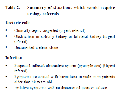
Initial treatment of ureteral colic After making the diagn osis, the next st ep in management is to identify those patients who require urgent intervention, in particular, urosepsis. These patients usually present with fever and chills. On physical examination, they may have flushing, septic-looks, tachycardia and even hypotension. As these patients can deteriorate rapidly, urgen t referral to casualty or urological care is important. Adequate fluid resuscitation, prompt and appropriate antibiotic treatment or urgent decompression of the system by either percutaneous nephrostomy or ureteric stent, is usually needed to control the sepsis. Bo th n o n - s te ro id a l an t i - in f l amma to ry d ru g s (NSAIDs) and opioids are effective and are recommended analgesics for pain control in patients with ureteric colic. Opioids are titratable and fast-acting, but they are more likely to cause nausea and vomiting. On the other hand, NSAIDs, which act directly on the pain mechanism pathway by inhibiting prostaglandin release, can cause gastrointestinal bleeding and renal impairment. Systemic review of the relative efficacy of NSAIDs and opioids revealed that both types of analgesics were effective in pain reduction. However, patients receiving opioids, especially pethidine, were more likely to have adverse events, e.g. vomiting. Therefore, NSAIDs are preferred unless there are contraindications. Pethidine is better avoided. Although up to two-thirds of ureteric stones will pass out spontaneously within four weeks from symptom onset, the interval between symptom onset and stone passage is highly variable. Therefore, these patients should be referred to specialty care for further monitoring and management of the stone. A tr ial of medic al exp ulsive therapy may be considered for these patients, especially for those with lower ureteric stones. Both alpha blockers (e.g. tamsulosin, terazosin, and doxazosin) and calcium channel blockers (e.g . nifedipine) have been investigated extensively for their role in facilitating ureteric stones passage. The mechanism of action is thought to be rel ated to their inh ibition on the u reter ic smo oth muscle spasm. Systemic review showed that a 2- to 4-week course of alpha blockers or calcium channel blo ckers was well-tolera ted. They could shor ten ston e pass ag e time and in cr ease s to ne e xp ul sion rate.9,10 Medical expulsive therapy is especially useful fo r ure te ri c s to nes o f mod er ate s ize (> 5mm b ut £ 10mm) at the distal end because stones less than 5mm can usually pass out spontaneously within four weeks even without any intervention. The use of alpha blockers has been shown to increase the stone passage rate by 29%.9 Prevention of stone recurrence Metabolic evaluation which include both blood (serum calcium, phosphate level, etc) and urine tests (both spot urine and 24 hour urine tests) is generally indicated only in those patients with recurrent stone formation.11 Another important investigation is stone analysis, as the identification of the stone composition will help to direct dietary advice and prophylactic measures. Therefore, we should always try to send any stone passed for chemical analysis whenever feasible. For those patients with no identifiable underlying cause, basic dietary advice should be given. Increased f lu id the r ap y is p e rh ap s th e c he ap es t and s af es t treatment and should be encouraged.12 Whil e the amount of fluid intake may not be easily translated into urine volume due to other routes of water loss, the general advice is to have at least 2 litres of urine passed every day or at least have sufficient fluid intake to maintain a clear coloured urine. But soft drinks (e.g. coke) have been shown to increase stone recurrence r a t e an d th e r e fo r e s h o u ld b e a v o i d e d . 13 Oth e r beverages, such as tea and coffee are less significant unless they are taken in excess. Dietary calcium restriction has now been shown to be ineffective, in fact it will increase stone recurrence rate instead.12 Current recommendation is to have a normal dietary calcium intake (about 800 to 1000 mg per day), unless the patient is shown to have absorptive hypercalciuria. Oxalate food should only be restricted when and if taken in excess. Reduced salt and animal protein intake is always advisable. The intake of citrus fruit is recommended, as it can increase urine pH and decrease stone recurrence. Urinary tract infection Lower urinary tract infection (LUTI) is probably one of the commonest urological conditions in the primary care setting. Lower urinary tract infection is defined as an infection confined to the bladder and urethral regions. Females have a much higher chance of getting an urinary tract infection (UTI) than males, with almost one-third of women having one episode of UTI requiring antibiotic therapy before the age of 24 and up to 50% of the female population suffering from at least one urinary tract infection in their lifetime.14 Diagnosis of simple cystitis is usually straightforward and relies heavily on clinical info rma t ion o bt a in e d f rom h i sto ry and p hy s i ca l examination. The classical presentations of lower urinary tract infection include dysuria, urinary frequency, urgency, haematuria, and suprapubic pain. Common precipitating events associated with LUTI are recent sexual activity, pregnancy, use of diaphragm with spermicide, etc.15 For simple LUTI, physical examination i s us ua lly un remarkabl e ex ce pt mi ld s up rapu bi c tenderness. However, hints for underlying problems may be identified during examination. A distended bladder may suggest a possible voiding dysfunction. In males, it is essential to identify phimosis and meatal stenosis, which are predisposing factors for recurrent infections. A rectal examination may also help to identify prostatic tenderness, which suggests acute prostatitis. Neurological examination may sometimes identify patients with voiding dysfunction secondary to neurological conditions. Investigation Apart from clinical information obtained from a focused history taking and physical examination, certain diagnostic adjuncts may help to establish the diagnosis. Urinalysis and urine culture are probably the commonest investigations done in the emergency department. Urine dipstick Urine dipstick for assessment of urinary tract infection involves the detection of leukocyte esterase activity and/or nitrite, which is produced by nitriteproducing pathogens, in particular Gram-negative bacteria. This is a rapid screening test to detect pyuria, and haematuria. The reported sensitivity of dipstick test for leukocyte esterase alone ranges from 48-86% and specificity varies from 17-93%.16 On the other hand, the dipstick nitrite test has a sensitivity of 18-81%, specificity of 87-100% and positive predictive value of 50-96%.17, 18 Meta-analysis showed that the combination of nitrite and leukocyte esterase tests increased the sensitivity at the expense of increased false positive results. However, combination of negative dipstick test for nitrite and leukocyte esterase shows a negative predictive value for urinary tract infection of 96.9% and a specificity of 98.7%.19 This signifies that, in the absence of both urine nitrite and leukocyte esterase activity, an urinary tract infection, is unlikely. Other findings from the dipstick test for urinary t r ac t in fe c t io n in c lud e p re s e n c e o f mic ro s c op i c haematuria, proteinuria, and a rise in the urine pH, but these findings are less specific as compared to the urine nitrite and leukocyte esterase in diagnosing urinary tract infection, and are usually not considered to be important in this setting. Cautions should also be taken when interpreting results of the dipstick test in patients with urinary catheters, and pregnant women in whom the accuracy is much lower. Urine culture Routine urine culture may not be necessary for s imp l e c y s t i t i s in y o u n g n o n -p r e g n an t f ema l e s . Howe v e r, in c omp l i c a t e d c o n d i t io ns s u c h as in children, males, during pregnancy and in patients suffering from recurrent cystitis, urine culture is still regarded as the gold standard for the diagnosis of a bacterial urinary tract infection. It is important to identify the underlying pathogens and guide subsequent investigations. It can also facilitate the differentiation of a re-infection from a relapse in the context of recurrent infections.
The traditional cut-off level of significant bacteriuria count of 105 colony forming units of bacteria per millilitre of urine is mainly for diagnosing upper tract infection and may not be applicable to cystitis. As most individuals tend to drink more fluid when experiencing cystitis symptoms, this fluid loading may result in reduced urinary bacterial count.20 Moreover, frequent voiding during cystitis can itself decrease the bacterial count in urine. Therefore, some authors suggest that a lower bacteriuria cut-off level of 103 uropathogens, should be adopted for diagnosing cystitis.21 Imaging and other investigations Further imaging following a simple cystitis in the female is usually not indicated. However, further studies are necessary to identify any predisposing cause of infection in children and male patients who have confirmed cystitis. In men, uncomplicated urinary tract infection is uncommon, and early evaluation should be advocated. Traditionally, intravenous urography was recommended to assess possible upper urinary tract obstruction and abnormality identified in plain film or ultrasonography. Apart from providing an anatomical assessment, it also gave functional evaluation, though involving the use of ionizing radiation and contrast media. Cu r r e n t e v i d e n c e s h ows t h a t c omb i n a t i o n o f ultrasonography with plain films is good enough and as accurate as intravevous urography in detecting important urological abnormalities in men, which is probably also a safer option. These imaging modalities, coupled with the additional information from urinary flow rate, seldom miss important urinary tract abnormalities.22 Any further investigation should also include a fasting blood sugar le vel to rule ou t und erlyin g d iabet es or gluco se intolerance. Antimicrobial therapy There is controversy over the optimal duration of antibiotic therapy. For young and non-pregnant females with acute uncomplicated cystitis, the preferred treatment is a three-day course of trimethoprim-sulfamethoxazole (Septrin) or quinolones, which are contraindicated in pregnancy.23 A longer course of therapy, usually 7-10 days, is recommended in diabetic women, during pregnancy, and patients with symptoms for more than 7 days or with other evidence of complicated urinary tract infections. However, in male patients with uncomplicated in f e c t io n , a mi n imum o f 7 d a y s o f t h e r a p y i s recommended as the presence of complicating factors is relatively more common. Recurrent cystitis When patients present with recurrent episodes of cystitis, more detailed investigations should be performed. Urine cultures during every attack of cystitis are essential for establishing the diagnosis, as well as aiding subsequent management. These should be carefully reviewed especially in patients with documented positive urine cultures in every, or the majority of attacks. If recurrent cystitis is caused by the same pathogen, this is highly suggestive of treatment failure or relapsing infection. Treatment failure can be due to resistant microbial strains, inappropriate antibiotics prescription or poor drug compliance. However, if there is no evidence to suggest treatment failure, the possibility of relapsing infection should be seriously considered. Relapse is often associated with urinary tract abnormalities, which are either structural or functional. The commonest causes include urolithiasis, vesico-ureteric reflux, presence of significant residual urine and bladder diverticulum. Therefore, these patients should be referred to the urologist for further evaluation. Unless the underlying cause is corrected, the pathogens may persist and these patients would continue to have recurrent episodes of infection. If the underlying complicating factors could not be corrected, long-term suppressive antibiotics therapy is needed to help to prevent frequent infections. Re-infection is diagnosed when patients have repeated positive urine cultures of different microorganisms. Possible causes for this condition include diabetes, poor hygiene, and immuno-compromised state. Treatment should be directed to rule out undiagnosed diabetes or an immune-compromised status. Patient education is vital. Patients should be instructed regarding the correct way of cleansing the perineum after micturition, the practice of post-coital voiding, and the importance of adequate fluid intake. However, for patients without any identifiable cause, a prolonged course of low dose prophylactic antibiotics (at least 6 months) should be considered. The choice of prophylactic antibiotics includes nitrofurantoin, tr imeth op rim/s ul famethox az ol e, or th e qu ino lo ne group, etc.23 However, there are patients who are labelled as suffering from ¡°recurrent cystitis¡± but never have had actual documented positive cultures. These patients should be referred to an urologist for further workup and investigation of the underlying irritating lower urinary tract symptoms. If haematuria is present, urgent referral may be necessary to rule out serious bladder pathology such as a tumour (the so called ¡°malignant cystitis¡±) or bladder stone, etc. Urine for cytology, plain radiography of a KUB view should also be performed to facilitate subsequent urological management. Key messages
Chi-fai Ng, MBChB (CUHK), FHKCS, FRCS (Ed) (Urol), FHKAM (Surg) Correspondence to: Professor Chi-fai Ng, Department of Surgery, Clinical Science Building, Prince of Wales Hospital, Shatin, Hong Kong SAR. Reference
|
|