|
March 2011, Volume 33, No. 1
|
Case Report
|
An 8 year-old-boy with fever, severe bilateral calf pain and toe-walkingJulie Y Chen 陳芸 HK Pract 2011;33:31-38 Summary Benign acute childhood myositis (BACM) is rare. It has been regarded as a dramatic complication of viral respiratory tract infection, particularly influenza B, whose clinical resolution is spontaneous, favourable and equally dramatic. This case report describes a clinical presentation which is consistent with the literature and offers an approach to ruling out sinister differential diagnoses. Awareness of this condition may help front-line clinicians to consider a selective approach to pursuing investigations. 摘要 良性急性兒童肌肉炎症(BACM)是病毒性呼吸道感染,尤其是乙型流感感染的併發症。它罕見而突發。它會自然康復,過程理想, 而同樣地令人感到突然。本報告敍述此病一案例的臨床表現,它和文獻上記載的很相近。同時提出一個與其他嚴重疾病辨症的方法。 對這種炎症的認知有助前線醫生為病人挑選適當的檢驗方案。 Introduction Upper respiratory tract infections (URTI) are common in the community. Both doctors and patients are well familiar with the usual litany of symptoms and signs as well as the natural history of the illness. One of the unusual and anxiety-provoking complications of URTI is benign acute childhood myositis (BACM), a self-limited muscle syndrome seen in school age children.1 It is more often associated with URTI due to influenza B 2 than with other viruses. It was originally described in 1957 as myalgia cruris epidemica 3 and is also known as influenza-associated myositis (IAM).3 Recent scattered case reports and case series have been reported from Australia,4 North America,5,6 the United Kingdom,7 India 8 and Taiwan,2 all describing a typical patient profile, presentation and clinical course. Awareness of the existence of this entity and its dramatic clinical presentation, particularly during an influenza season, would help clinicians recognize a benign self-limited condition and may pre-empt hospital admission and excessive testing for serious disease.
The following case was encountered in a non-clinical setting, and is meant to
highlight distinctive clinical features which may alert the clinician to the diagnosis
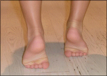
of BACM, rather than to serve as an exemplar for practice. Case presentation BB*, an 8-year-old Eurasian Hong Kong boy, presented with bilateral calf pain and fever while on holiday in Thailand in February 2010. Before leaving on his trip, he had a fever of 39oC (responsive to ibuprofen), runny nose, cough, myalgia and general malaise for 3 days with the fever resolving on day 4. Upon arrival in Thailand on day 5, he was asymptomatic but developed a low grade fever later the same evening, coupled with a complaint of sore calves. On the morning of day 6, he had alarmed his parents because he was unable to get out of bed to use the toilet. He preferred to crawl on all fours or to ambulate upright by "toe-walking" (distinct from "tip-toeing") (Figure 1) stating that his calves hurt when he tried to stand or walk. He said he felt no discomfort moving around as long as he was not weight bearing. His appetite and energy level were unaffected with his only complaint being that he wished he could join his friends in the pool. Figure 1: Toe-walking
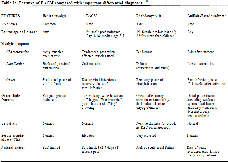
When seen in his hotel room that day, BB appeared well, and was actively playing computer games. He was afebrile. Both lower extremities were of normal colour and temperature without evidence of swelling or skin rash. There was bilateral calf tenderness and calf pain with passive dorsiflexion of the ankle bilaterally. Muscle tone, power, sensation and tendon reflexes were grossly normal and symmetrical. Additionally, the urine was observed to be pale yellow in colour. BB had previously been well with no similar episodes of calf pain in the past. His past medical history was unremarkable for chronic conditions or major medical illnesses. Routine childhood immunizations were up-to-date. He had not received the influenza vaccination this year. He was typically active in sporting activities but had not reported any trauma or undue exertion prior to his illness. There was no family history of neuromuscular disease. After 36 hours of rest and enthusiastic use of a wheelchair to ambulate, BB suddenly started walking normally, with complete resolution of the calf pain, and was engaging in all holiday activities. His return to Hong Kong was uneventful and he continues to remain well. Discussion This diagnosis of BACM was made solely on clinical grounds and based on a review of published available medical literature accessible on the internet, as laboratory investigations were not readily available in this particular setting. A subsequent, more comprehensive review of the literature revealed that there was a paucity of information about BACM, with this mostly in the form of case reports or short textbook entries. Typical clinical picture A textbook presentation of BACM would have a school aged boy presenting with bilateral calf pain and compensatory gait in the face of a resolving URTI during influenza season. The finding of symmetric calf pain relates to the very typical involvement of gastrocnemius and soleus muscles although very rarely the thighs or upper extremities may be affected. The patient may refuse to walk (crawling or "bottom shuffling")4 or use a characteristic wide-based and straight-legged "Frankenstein"6 gait or "toe-walking" 1 to avoid stretching the calf muscles. Aside from very tender calf muscles, the patient would have a normal neurologic examination (sensory, motor and deep tendon reflexes) and appear systemically well. Laboratory findings There is marked elevation of serum creatine kinase (CK) in BACM, usually up to 2 to 50 times normal (median 4100 IU/L),3 normal CK being up to 240 IU/L.4 These values return to normal within a month.1 Often there is also leukopenia and an elevation of liver transaminases.6 Epidemiology and aetiology The actual incidence and prevalence of the condition is not known. Though this is a rare condition documented only by case reports, it seems that among children with influenza, the prevalence is quite high. In a retrospective hospital-based study of 197 Taiwanese children with culture-confirmed influenza, 46 (23%) were identified with BACM.2 Boys are affected more frequently than girls by a ratio of 2:1 3 leading to the question of whether there was a genetic association with this condition. The evidence linking BACM to a viral cause, specifically influenza B, is fairly consistent with the McIntyre and Doherty 11 study. Among 77 BACM patients studied 62% were test positive for influenza B and 25% for influenza A. Other entities have also been associated with BACM including parainfluenza, adenovirus, rotavirus, and mycoplasma. In the case of BB, the aetiologic agent was likely influenza B, as the swine and seasonal flu monitor published by the Hong Kong Centre for Health Protection indicated that during the time of BB's initial illness in Hong Kong, the predominant circulating strain was influenza B, though the overall seasonal influenza activity was low.12 There have been sporadic reports of BACM in the literature mostly occurring during late winter and early spring, consistent with the timing of outbreaks of viral respiratory illness in the northern hemisphere. Pathogenesis BACM is considered a myositis as defined by consistently elevated serum CK and characteristic pathologic findings on muscle biopsies.5 The exact pathogenesis is unclear but it has been postulated to involve a post infectious autoimmune mechanism (supported by the diversity of associated agents and the appearance of muscular pain after other symptoms subside) caused by direct invasion by viral particles (as seen on muscle biopsy).13 Differential diagnoses Despite a suggestive clinical picture, key differential diagnoses, should be considered including Guillain-Barré syndrome 14 and rhabdomyolysis 9 both of which could have potentially serious outcomes but both of which also exhibit distinguishing features. A comparison of the features of these two conditions with BACM and benign viral myalgia is presented in Table 1.
Guillain-Barré syndrome is a peripheral neuropathy with a varied spectrum of subtypes but whose common hallmark is a clinical picture of rapidly progressive limb weakness peaking within four weeks, associated with a history of preceding respiratory or gastrointestinal infection.14 Other possible neurologic findings include loss of the deep tendon reflexes, sensory symptoms (e.g. paraesthesia), autonomic signs (e.g. tachycardia), and cranial nerve involvement (e.g. facial weakness). A child's refusal to walk or to weight-bear may be due to a neurological deficit, i.e. lower extremity weakness, or may be a protective reaction, i.e. to avoid muscle pain 7 which may be a difficult but important differentiating feature, as BACM patients are neurologically intact. Rhabdomyolysis is positioned on the severe end of the spectrum of myositis entities and is of particular concern as it may lead to life-threatening complications such as myoglobin-induced acute renal failure, compartment syndrome and electrolyte disturbances.3 Contents of damaged skeletal muscle cells, including the haem protein myoglobin and the enzyme creatine kinase (CK), are released into the systemic circulation and are diagnostic (by definition over 1000 IU/L9 and may rise to 500,000 IU/L10) markers for the illness. Urine appears reddish brown as myoglobin is filtered into the urine, and will also dip positive for blood (haem) due to the cross reactivity of haem and myoglobin. However, no red blood cells should be seen under microscopic examination of the urine. Epidemiologically, rhabdomyolysis has been reported more frequently in girls and has been associated with influenza A 3 which in turn has been linked with causing more severe disease 2. Prognosis The prognosis for BACM is excellent, with spontaneous, rapid and full recovery within 3-7 days.1 Only rest, analgesia and supportive measures are required. In terms of the risk of developing rhabdomyolysis, Agyeman et al3 described a series of 316 clinically similar cases which they termed influenza-associated myositis in children. Of these, 10 (3%) developed rhabdomyolysis with 8 patients going into acute renal failure. A study of rhabdomyolysis in a series of paediatric patients by Mannix et al15 found that 38% were due to a viral myositis and of those, influenza was the most common aetiologic agent. Recovered patients do not seem to be any increased risk for recurrence although it may happen, usually in the context of a first exposure to a different virus.1 Although vaccination is believed to reduce the risk of common complications such as otitis media and pneumonia, it is not known if it affects the likelihood of developing BACM.6 Proposed diagnostic approach (Figure 2) Figure 2 : Summary of a proposed diagnostic approach to an otherwise healthy child presenting with sudden difficulty walking, bilateral calf pain, and URTI
Recognizing the stereotypical clinical presentation and biochemical features of BACM could prevent unnecessary invasive tests. On the other hand, BACM must be differentiated from conditions with serious consequences so erring on the side of caution by extensive testing is understandable. It would be reasonable to manage a patient in whom there was a strong clinical suspicion for BACM as an outpatient in a primary care setting where laboratory testing and results were rapidly available and follow up assured. This would enable monitoring for rhabdomyolysis or following up a mildly elevated CK which may reflect early Guillane-Barré syndrome. In Hong Kong this would probably mean the Accident and Emergency Department (AED) or similarly resourced outpatient setting. In their series of 32 cases seen at a university based primary care paediatric clinic, Zafeirou et al16 indicated that the majority of patients were evaluated on an outpatient basis with blood and urine tests and then followed up at a subspecialist outpatient clinic. Rennie et al7 suggested that BACM could be evaluated appropriately in an accident and emergency department if adequate follow up arrangements can be made and this is echoed by King et al4 who further stressed the importance of ensuring close clinical follow up. Non-classic presentation of BACM would trigger a more urgent referral for more comprehensive assessment of serious differentials. Possible red flags are outlined in the algorithm. Conclusion In a child presenting with bilateral calf pain and sudden difficulty walking in the setting of a viral illness, it remains critical to exclude the possible sinister causes but to also recognize BACM as an uncommon but dramatic complication of a common viral infection. A possible diagnostic approach is thus proposed to help clinicians address parental anxiety and to facilitate the outpatient assessment of patients with this condition. Acknowledgement I would like to gratefully knowledge the cooperation and the encouragement of BB and his parents, in the compilation of this case report.
Julie Y Chen, MD, FCFPC
Assistant Professor, Department of Family Medicine and Primary Care and Institute of Medical and Health Sciences Education, The University of Hong Kong. Correspondence to : Dr Julie Y Chen, Department of Family Medicine and Primary Care, 3/F Ap Lei Chau Clinic, 161 Main Street, Ap Lei Chau, Hong Kong SAR.
References
|
|


